 Back
BackCell Structure and Function, and Microbial Metabolism: Key Diagrams and Processes
Study Guide - Smart Notes
 Tailored notes based on your materials, expanded with key definitions, examples, and context.
Cell Structure and Function
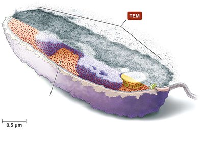
Prokaryotic Cell Structure
Prokaryotic cells, such as bacteria, are structurally simpler than eukaryotic cells. They lack membrane-bound organelles and a true nucleus, but possess specialized structures for survival and adaptation.
Cytoplasmic Membrane: A phospholipid bilayer that controls the movement of substances in and out of the cell.
Cell Wall: Provides structural support and shape; composed mainly of peptidoglycan in bacteria.
Glycocalyx: An external layer that can be a capsule or slime layer, aiding in protection and adherence.
Nucleoid: Region containing the cell's genetic material (DNA), not surrounded by a membrane.
Ribosomes: Sites of protein synthesis, smaller (70S) than those in eukaryotes.
Flagellum: Used for motility; structure varies between Gram-positive and Gram-negative bacteria.
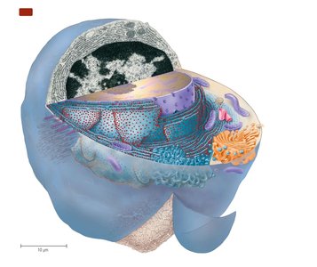
Eukaryotic Cell Structure
Eukaryotic cells are more complex, containing membrane-bound organelles and a defined nucleus. These features allow compartmentalization of cellular processes.
Nucleus: Contains genetic material and is surrounded by a nuclear envelope with pores.
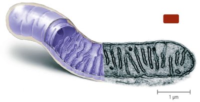
Mitochondria: Sites of aerobic respiration and ATP production.
Endoplasmic Reticulum (ER): Rough ER is studded with ribosomes for protein synthesis; smooth ER is involved in lipid synthesis.
Golgi Body: Modifies, sorts, and packages proteins and lipids for secretion or delivery to other organelles.
Lysosomes: Contain digestive enzymes for breakdown of macromolecules.
Cytoskeleton: Provides structural support and facilitates movement.
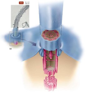
Cilia and Flagella: Used for movement; have a characteristic "9+2" arrangement of microtubules.
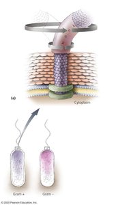
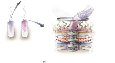
Bacterial Flagella Structure and Function
Bacterial flagella are complex structures that enable motility. Their arrangement and structure differ between Gram-positive and Gram-negative bacteria.
Filament: The long, whip-like part composed of flagellin protein.
Hook: Connects the filament to the basal body.
Basal Body: Anchors the flagellum to the cell wall and membrane; structure varies with cell wall type.
Rotation: Flagella rotate like propellers, enabling movement toward or away from stimuli (chemotaxis).
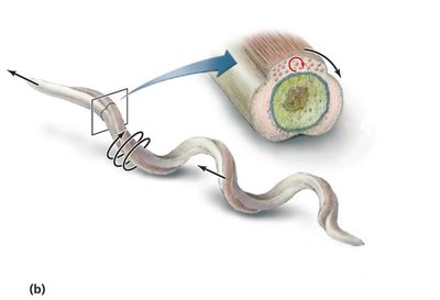
Axial Filaments in Spirochetes
Spirochetes possess unique flagella called axial filaments or endoflagella, located in the periplasmic space. Their rotation causes the entire cell to move in a corkscrew motion, aiding in movement through viscous environments.
Axial Filament: Composed of endoflagella wrapped around the cell body.
Movement: Rotation of the axial filament propels the spirochete forward.
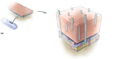
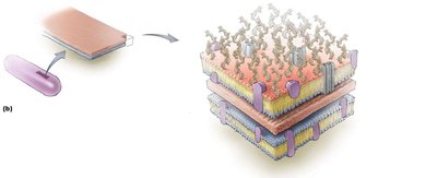
Gram-Positive and Gram-Negative Cell Walls
The cell wall structure is a key feature distinguishing Gram-positive from Gram-negative bacteria, affecting staining properties and susceptibility to antibiotics.
Gram-Positive: Thick peptidoglycan layer, teichoic acids, no outer membrane.
Gram-Negative: Thin peptidoglycan layer, outer membrane with lipopolysaccharide (LPS), periplasmic space.
Feature | Gram-Positive | Gram-Negative |
|---|---|---|
Peptidoglycan | Thick | Thin |
Teichoic acids | Present | Absent |
Outer membrane | Absent | Present |
LPS | Absent | Present |
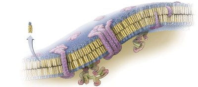
Prokaryotic Cytoplasmic Membrane
The cytoplasmic membrane is a selectively permeable barrier composed of a phospholipid bilayer with embedded proteins. It regulates the passage of substances and is involved in energy generation and cell signaling.
Phospholipid Bilayer: Hydrophilic heads face outward, hydrophobic tails inward.
Integral and Peripheral Proteins: Serve as channels, carriers, receptors, and enzymes.
Eukaryotic Flagella and Cilia
Eukaryotic flagella and cilia are composed of microtubules arranged in a "9+2" pattern. The basal body anchors these structures to the cell, and their coordinated movement enables locomotion or movement of substances across the cell surface.
Flagella: Longer, usually one or a few per cell, move in a whip-like fashion.
Cilia: Shorter, numerous, move in coordinated waves.
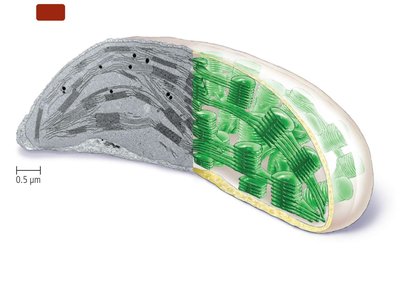
Mitochondria and Chloroplasts
Mitochondria and chloroplasts are double-membraned organelles involved in energy conversion. Mitochondria are the site of aerobic respiration, while chloroplasts carry out photosynthesis in plants and algae.
Mitochondria: Contain cristae (folded inner membrane) for increased surface area for ATP production.
Chloroplasts: Contain thylakoids stacked into grana, where light-dependent reactions of photosynthesis occur.
Microbial Metabolism
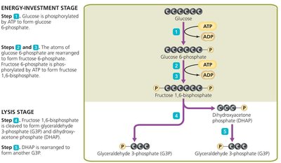
Glycolysis (Embden-Meyerhof-Parnas Pathway)
Glycolysis is the metabolic pathway that converts glucose into pyruvate, generating ATP and NADH. It occurs in the cytoplasm and is the first step in both aerobic and anaerobic respiration.
Energy-Investment Stage: 2 ATP are used to phosphorylate glucose and its intermediates.
Lysis Stage: 6-carbon sugar is split into two 3-carbon molecules.
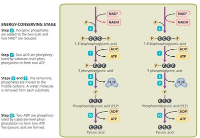
Energy-Conserving Stage: 4 ATP and 2 NADH are produced per glucose molecule.
Net Reaction:
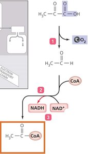
Pyruvate Dehydrogenase Complex
After glycolysis, pyruvate is converted to acetyl-CoA by the pyruvate dehydrogenase complex. This reaction links glycolysis to the citric acid cycle and produces NADH and CO2.
Decarboxylation: Pyruvate loses a carbon as CO2.
Oxidation: Remaining 2-carbon fragment is oxidized, reducing NAD+ to NADH.
Coenzyme A Addition: Forms acetyl-CoA, which enters the citric acid cycle.
Reaction:
Citric Acid Cycle (Krebs Cycle)
The citric acid cycle is a series of enzyme-catalyzed reactions in the mitochondrial matrix (or cytoplasm of prokaryotes) that oxidize acetyl-CoA to CO2, generating NADH, FADH2, and GTP/ATP.
Acetyl-CoA combines with oxaloacetate to form citrate.
Through a series of steps, citrate is oxidized, releasing CO2 and transferring electrons to NAD+ and FAD.
Products per turn: 3 NADH, 1 FADH2, 1 GTP (or ATP), 2 CO2.
Overall Reaction (per glucose):
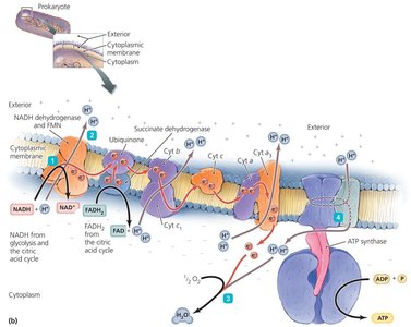
Electron Transport Chain (ETC)
The electron transport chain is a series of protein complexes located in the inner mitochondrial membrane (eukaryotes) or plasma membrane (prokaryotes). Electrons from NADH and FADH2 are transferred through the chain, driving proton pumping and ATP synthesis.
Electron Carriers: NADH, FADH2, ubiquinone, cytochromes.
Proton Gradient: Electron transfer pumps protons across the membrane, creating an electrochemical gradient.
ATP Synthase: Protons flow back through ATP synthase, driving ATP production from ADP and Pi.
Oxygen: Final electron acceptor in aerobic respiration, forming water.
Overall Reaction:
Calvin-Benson Cycle
The Calvin-Benson cycle is the set of light-independent reactions in photosynthesis that fix carbon dioxide into organic molecules. It occurs in the stroma of chloroplasts.
Key Steps: Carbon fixation, reduction, and regeneration of ribulose bisphosphate (RuBP).
Products: Glyceraldehyde 3-phosphate (G3P), which can be used to form glucose and other carbohydrates.
Energy Input: ATP and NADPH from the light-dependent reactions are consumed.
Overall Reaction:
