 Back
BackCell Structure and Function: Prokaryotes vs. Eukaryotes
Study Guide - Smart Notes
 Tailored notes based on your materials, expanded with key definitions, examples, and context.
Cell Structure: Prokaryotes vs. Eukaryotes
Overview of Cell Types
Cells are classified into two main types: prokaryotic and eukaryotic. Prokaryotes include bacteria and archaea, while eukaryotes encompass animals, plants, fungi, and protists. Understanding their structural differences is fundamental in microbiology.
Prokaryotes: Lack a nucleus and membrane-bound organelles; DNA is located in the nucleoid.
Eukaryotes: Possess a nucleus and various membrane-bound organelles.
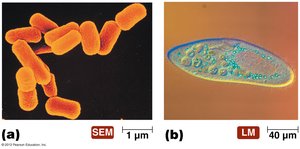
Size: Prokaryotes are generally smaller (1-10 µm) than eukaryotes (10-100 µm).
Examples: Escherichia coli (prokaryote), Paramecium (eukaryote).
External Structures of Prokaryotic Cells
Appendages: Flagella, Pili, and Fimbriae
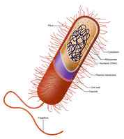
Prokaryotic cells possess various external structures that aid in movement, attachment, and genetic exchange.
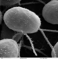
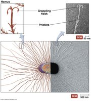
Flagella: Long, whip-like structures used for locomotion. Originate from the cell membrane and rotate like a propeller.
Pili: Rigid, hollow tubes made of pilin protein. Function in attachment and transfer of DNA (conjugation).
Fimbriae: Short, hair-like fibers involved in attachment to surfaces and biofilm formation.
Flagella Arrangements and Motility
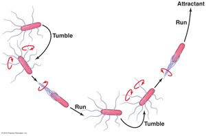
Flagella can be arranged in different patterns, affecting bacterial movement. Motility involves alternating between 'runs' and 'tumbles' to navigate toward attractants.
Monotrichous: Single flagellum at one end.
Lophotrichous: Multiple flagella at one end.
Amphitrichous: Flagella at both ends.
Peritrichous: Flagella distributed over the entire cell surface.
Movement: Bacteria alternate between straight runs and random tumbles.
Special Flagella: Axial Filaments (Endoflagella)
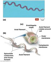
Some bacteria, especially spirochetes, possess axial filaments that run along the periplasmic space, enabling corkscrew-like movement.
Axial Filament: Originates in the cell membrane, rotates to move the cell.
Associated with: Spirochetes (e.g., Treponema).
Glycocalyx: Slime Layer and Capsule
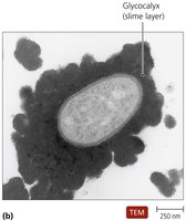
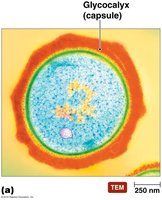
The glycocalyx is a protective coating outside the cell wall, which can be either a slime layer or a capsule.
Slime Layer: Loosely attached, composed of polysaccharide fibers. Functions in protection and attachment, often associated with biofilms.
Capsule: Firmly attached, composed of polysaccharides or polypeptides. Protects against phagocytosis, acts as an osmotic barrier, and is a virulence factor.
Cell Envelope: Cell Wall and Membrane
Structure and Function of the Cell Wall
The cell wall provides structural support, determines cell shape, and protects against lysis. It is a major target for antibiotics.
Peptidoglycan: Main component of bacterial cell walls.
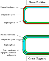
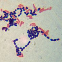
Gram-Positive: Thick peptidoglycan layer, contains teichoic acids, stains purple.
Gram-Negative: Thin peptidoglycan layer, outer membrane with LPS, stains pink.
Gram Staining and Cell Wall Differences
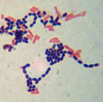
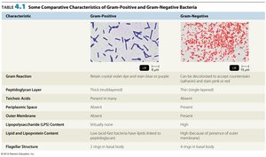
Gram staining differentiates bacteria based on cell wall structure. This distinction is crucial for diagnosis and treatment.
Gram-Positive: Thick peptidoglycan, teichoic acids, no outer membrane.
Gram-Negative: Thin peptidoglycan, outer membrane with LPS, porins, periplasmic space.
Characteristic | Gram-Positive | Gram-Negative |
|---|---|---|
Gram Reaction | Retain crystal violet, stain blue/purple | Lose crystal violet, stain red/pink |
Peptidoglycan Layer | Thick, multilayered | Thin, single-layered |
Teichoic Acid | Present | Absent |
Outer Membrane | Absent | Present |
LPS Content | Low/none | High |
Flagella Structure | 2 rings in basal body | 4 rings in basal body |
Cytoplasmic Membrane
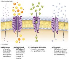
The cytoplasmic membrane is a selectively permeable barrier composed of a phospholipid bilayer and proteins. It regulates transport, energy reactions, and secretion.
Phospholipid Bilayer: Hydrophilic heads and hydrophobic tails.
Integral and Peripheral Proteins: Facilitate transport and cellular functions.
Transport Processes: Diffusion, facilitated diffusion, osmosis, active transport, group translocation.
Internal Structures of Prokaryotic Cells
Cytoplasm and Inclusions
The cytoplasm is a gelatinous solution containing water, enzymes, ribosomes, DNA, and inclusion bodies. Inclusions store nutrients and other substances.
Cytosol: Liquid portion of cytoplasm.
Inclusions: Reserve deposits of chemicals (e.g., glycogen, polyphosphate).
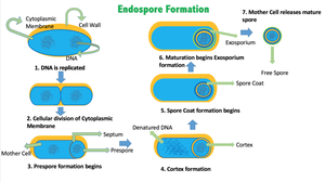
Endospores: Defensive structures produced by some bacteria under stress.
Nucleoid and Chromosome
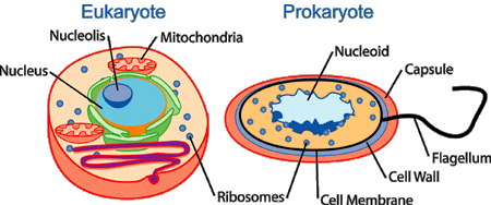
The nucleoid is the region where the bacterial chromosome (single, circular DNA) is located. It is not membrane-bound.
Chromosome: Contains essential genetic information.
Nucleoid: Dense area of DNA aggregation.
Ribosomes
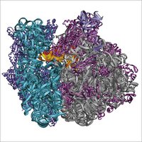
Bacterial ribosomes are composed of RNA and protein, and are smaller (70S) than eukaryotic ribosomes (80S). They are the site of protein synthesis.
Structure: 70S, made of 50S and 30S subunits.
Function: Translate mRNA into protein.
Plasmids
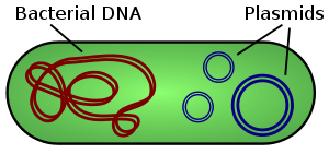
Plasmids are small, circular, non-essential DNA molecules found in the cytoplasm. They often carry genes for antibiotic resistance or toxin production.
Function: Confer protective traits, easily transferred between bacteria.
Rare in eukaryotes.
Cytoskeleton
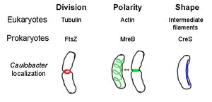
The prokaryotic cytoskeleton is composed of protein fibers that help maintain cell shape, assist in cell division, and segregate DNA molecules.
Proteins: FtsZ, MreB, CreS.
Functions: Cell division, polarity, shape.
Structural Characteristics of Prokaryotes
Comparison Table: Archaea vs. Bacteria
Prokaryotes are divided into two domains: Archaea and Bacteria. They share some features but differ in cell wall composition, membrane lipids, and other structures.
Feature | Archaea | Bacteria |
|---|---|---|
Glycocalyx | Polysaccharide or polypeptide | Polysaccharide or polypeptide |
Flagella | Present in some, rotate as bundles | Present in some, rotate as bundles |
Pili | None discovered | Present, used for attachment and DNA exchange |
Cell Wall | Present in most, composed of protein or polysaccharide | Present in most, composed of peptidoglycan |
Cytoplasmic Membrane | Ether-linked lipids | Ester-linked lipids |
Cytoplasm | Contains circular DNA, 70S ribosomes | Contains circular DNA, 70S ribosomes |
Eukaryotic Cell Structure
Typical Eukaryotic Cell
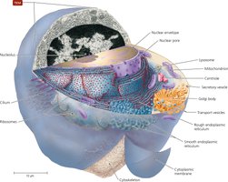
Eukaryotic cells are more complex, containing a nucleus and various organelles. They have a cytoplasmic membrane, cell wall (in some), and external structures like flagella and cilia.
Nucleus: Contains DNA, surrounded by nuclear envelope.
Organelles: Mitochondria, endoplasmic reticulum, Golgi body, lysosomes, peroxisomes, vacuoles.
Flagella and Cilia: Used for movement, structurally different from prokaryotic flagella.
External Structures of Eukaryotic Cells
Eukaryotic cells may have a glycocalyx, which is less organized than prokaryotic capsules. It aids in cell-to-cell recognition and protection.
Glycocalyx: Anchors cells, strengthens surface, prevents dehydration.
Cell Wall: Present in fungi, algae, plants, some protozoa.
Eukaryotic Cytoplasmic Membrane
The cytoplasmic membrane is a fluid mosaic of phospholipids and proteins, containing steroid lipids for fluidity and membrane rafts for specialized functions.
Functions: Controls movement, maintains cell integrity.
Flagella and Cilia in Eukaryotes
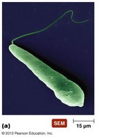
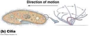
Eukaryotic flagella are composed of microtubules and move by undulation, not rotation. Cilia are shorter, more numerous, and move substances across the cell surface.
Flagella: Tubulin-based, anchored by basal body.
Cilia: Coordinated beating for movement and substance transport.
Nonmembranous Organelles
Eukaryotic ribosomes are larger (80S) and cytoskeleton is more extensive, composed of microtubules, microfilaments, and intermediate filaments.
Ribosomes: 80S, composed of 60S and 40S subunits.
Cytoskeleton: Anchors organelles, shapes cell.
Membranous Organelles
Eukaryotic cells contain several membrane-bound organelles, each with specialized functions.
Nucleus: Contains DNA, nucleoplasm, nucleoli.
Endoplasmic Reticulum (ER): Smooth (SER) and rough (RER), involved in transport and synthesis.
Golgi Body: Processes and packages molecules for export.
Lysosomes, Peroxisomes, Vacuoles: Store and transfer chemicals, digest macromolecules, degrade waste.
Mitochondria: Energy production (ATP), contains DNA and 70S ribosomes.
Chloroplasts: Photosynthesis, contains DNA and 70S ribosomes.
Endosymbiotic Theory
Origin of Eukaryotic Organelles
The endosymbiotic theory proposes that eukaryotes evolved from a symbiotic relationship between aerobic prokaryotes and larger anaerobic prokaryotes. Mitochondria and chloroplasts are believed to have originated from these internalized prokaryotes.
Mitochondria: Derived from aerobic prokaryotes.
Chloroplasts: Derived from photosynthetic prokaryotes.
Summary and Take Home Points
Know the major prokaryotic structures and their functions.
Know the major eukaryotic structures and their functions.
Be able to compare and contrast structures between eukaryotic and prokaryotic cells.
