 Back
BackCell Structure and Function: Prokaryotic and Eukaryotic Cells
Study Guide - Smart Notes
 Tailored notes based on your materials, expanded with key definitions, examples, and context.
Prokaryotic and Eukaryotic Cells: An Overview
Definitions and Key Differences
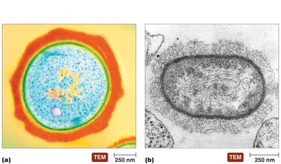
Cells are the fundamental units of life, classified into two main types: prokaryotic and eukaryotic. Understanding their structural differences is essential for microbiology.
Prokaryotes: Include bacteria and archaea. They lack a nucleus and membrane-bound organelles, and are typically 1.0–2.0 µm in diameter.
Eukaryotes: Include algae, protozoa, fungi, animals, and plants. They possess a nucleus and internal membrane-bound organelles, and are generally 10–100 µm in diameter.
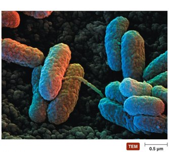
Bacterial Shapes and Arrangements
Bacteria exhibit a variety of shapes and arrangements, which are important for identification and classification.
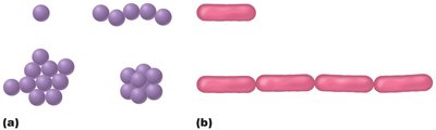
Cocci: Spherical bacteria, found singly, in chains (strepto-), clusters (staphylo-), or packets (sarcina).
Bacilli: Rod-shaped bacteria, found singly or in chains (strepto-).
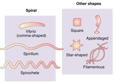
Other shapes include spiral forms (vibrio, spirillum, spirochete) and less common shapes such as square, star-shaped, appendaged, and filamentous.
External Structures of Bacterial Cells
Glycocalyces
The glycocalyx is a sticky, gelatinous layer surrounding the cell wall, which can be a capsule or a slime layer. It provides protection, aids in adherence, and contributes to pathogenicity.
Capsule: Organized, firmly attached glycocalyx; protects against phagocytosis.
Slime layer: Unorganized, loosely attached; aids in biofilm formation.
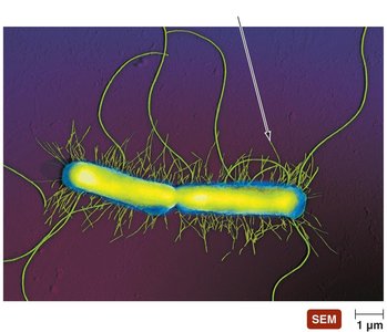
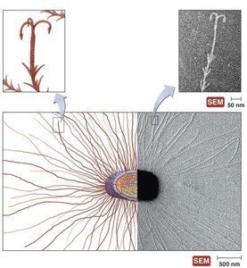
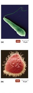
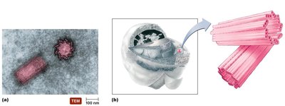
Flagella
Flagella are long, whip-like structures used for motility. Their structure differs between Gram-positive and Gram-negative bacteria, but the function is similar: propulsion.
Composed of filament, hook, and basal body.
Rotation of flagella enables movement toward or away from stimuli (taxis).
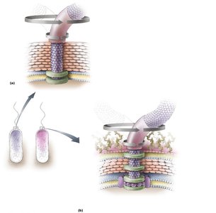
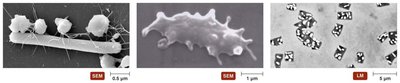
Fimbriae and Pili
Fimbriae are short, hair-like appendages for attachment, while pili (singular: pilus) are longer and involved in conjugation (transfer of genetic material).
Fimbriae: Enable adherence to surfaces and other cells.
Pili: Facilitate horizontal gene transfer via conjugation.
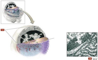
Bacterial Cell Walls
Structure and Function
The cell wall provides structural support, shape, and protection from osmotic forces. It is primarily composed of peptidoglycan, a mesh-like polymer of sugars and amino acids.
Resists antimicrobial drugs, but is targeted by many antibiotics.
Two main types: Gram-positive and Gram-negative.
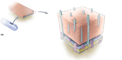
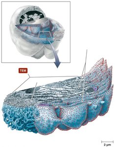
Gram-Positive Cell Walls
Gram-positive bacteria have thick peptidoglycan layers and contain teichoic acids, which contribute to cell wall stability and function.
Thick peptidoglycan layer.
Teichoic and lipoteichoic acids present.
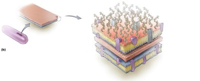
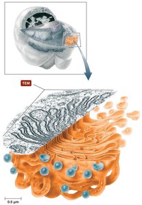
Gram-Negative Cell Walls
Gram-negative bacteria have a thin peptidoglycan layer and an outer membrane containing lipopolysaccharides (LPS), which includes lipid A (an endotoxin).
Thin peptidoglycan layer.
Outer membrane with LPS, porins, and periplasmic space.
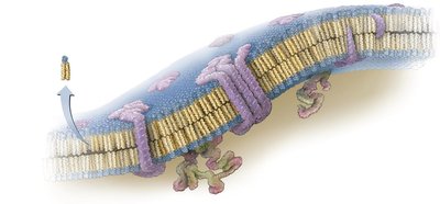
Bacterial Cytoplasmic Membranes
Structure and Function
The cytoplasmic membrane is a phospholipid bilayer with embedded proteins, serving as a selective barrier and site for metabolic activities.
Phospholipid heads are hydrophilic; tails are hydrophobic.
Integral and peripheral proteins facilitate transport and communication.
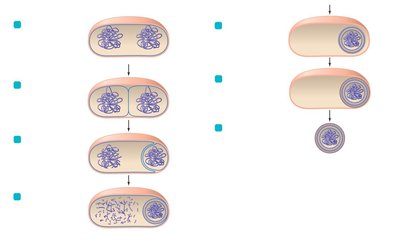
Cytoplasm of Bacteria
Cytosol, Inclusions, and Endospores
The cytoplasm contains the cytosol (mostly water), inclusions (storage granules), and the nucleoid (region with DNA). Some bacteria form endospores, highly resistant structures for survival under harsh conditions.
Cytosol: Liquid portion, contains DNA and metabolic enzymes.
Inclusions: Storage of nutrients (glycogen, sulfur, etc.).
Endospores: Dormant, resistant to heat, chemicals, and radiation.
External Structures of Archaea
Glycocalyces, Flagella, Fimbriae, and Hami
Archaea possess unique external structures, including glycocalyces, flagella, fimbriae, and hami. Hami are specialized, grappling hook-like appendages for attachment.
Glycocalyces: Aid in biofilm formation and adherence.
Flagella: Structurally distinct from bacterial flagella.
Fimbriae and hami: Attachment to surfaces.
Structure of Eukaryotic Cells
Glycocalyces, Cell Walls, and Cytoplasmic Membranes
Eukaryotic cells may have a glycocalyx, cell wall, and cytoplasmic membrane. The glycocalyx is less organized than in prokaryotes and aids in cell recognition and protection.
Cell walls: Found in plants, fungi, and some protists; composed of cellulose, chitin, or agar.
Cytoplasmic membrane: Similar phospholipid bilayer as prokaryotes.
Cytoplasm of Eukaryotes
Flagella, Cilia, and Nonmembranous Organelles
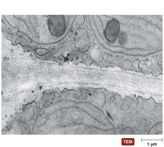
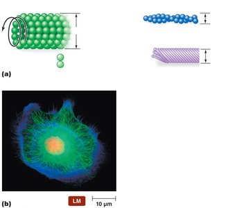
Eukaryotic cells contain complex cytoskeletal elements and organelles. Flagella and cilia are used for movement, while the cytoskeleton provides structural support.
Flagella: Long, whip-like; cilia: short, numerous.
Cytoskeleton: Microtubules, microfilaments, intermediate filaments.
Centrosome: Organizes microtubules.
Membranous Organelles
Eukaryotic cells contain membrane-bound organelles, each with specialized functions.
Nucleus: Contains genetic material (DNA).
Endoplasmic reticulum (ER): Rough ER (protein synthesis), Smooth ER (lipid synthesis).
Golgi body: Modifies, sorts, and packages proteins.
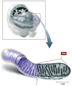
Mitochondria: Site of aerobic respiration; contains its own DNA.
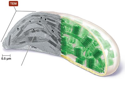
Chloroplasts: Site of photosynthesis in plants and algae.
Endosymbiotic Theory
Origin of Eukaryotic Organelles
The endosymbiotic theory proposes that eukaryotes originated from a symbiotic relationship between small aerobic prokaryotes and larger anaerobic prokaryotes. The smaller prokaryotes became mitochondria and chloroplasts.
Evidence: Mitochondria and chloroplasts have their own DNA, double membranes, and reproduce independently.
Significance: Explains the origin of key eukaryotic organelles.
----------------------------------------
