 Back
Cell Structure and Function: Prokaryotic and Eukaryotic Cells
Study Guide - Smart Notes
 Tailored notes based on your materials, expanded with key definitions, examples, and context.
Cell Structure and Function
Characteristics of Life
To be considered alive, an organism must exhibit several key characteristics: growth, reproduction, responsiveness, metabolism, and cellular structure. Viruses and prions challenge these definitions, as they lack some or all of these features.
Growth: Increase in size; occurs in all living cells but not in viruses.
Reproduction: Ability to reproduce independently; viruses require host cells, prions do not reproduce in the traditional sense.
Responsiveness: Ability to react to environmental stimuli; present in cells, limited or absent in viruses and prions.
Metabolism: Chemical reactions for energy; absent in viruses and prions.
Cellular Structure: Cells are the basic unit of life; viruses and prions lack cellular structure.

Overview of Prokaryotic and Eukaryotic Cells
Cells are the fundamental units of life. Organisms may be unicellular or multicellular. Cells are classified as either prokaryotic or eukaryotic based on their structural features.
Prokaryotes: Domains Bacteria and Archaea; lack a membrane-bound nucleus; typically smaller (≤1 µm).
Eukaryotes: Have a true nucleus and membrane-bound organelles; typically larger (10–100 µm).
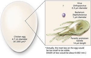
Cell Types and Structures
All living things are made up of cells. Prokaryotic and eukaryotic cells differ in complexity and internal organization.
Prokaryotic Cells: Structurally simple, no nucleus, unicellular, lack internal compartmentalization.
Eukaryotic Cells: Structurally complex, contain a nucleus, and possess various organelles.
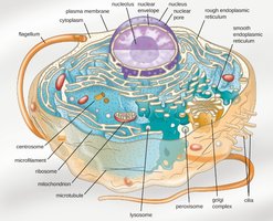
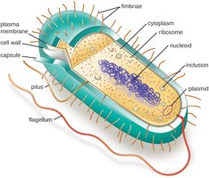
Prokaryotic Cell Structure
Glycocalyx (Capsule)
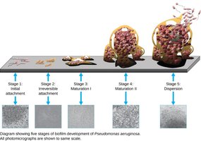
The glycocalyx is a sugar-rich, gelatinous layer outside some prokaryotic cells. When tightly bound, it is called a capsule.
Functions: Source of nutrition, aids in biofilm formation, and acts as a virulence factor by protecting cells from host defenses.
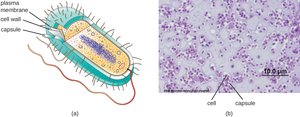
Flagella
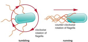
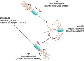
Flagella are long, whip-like appendages used for motility. Their arrangement varies among species and can be used for classification.
Function: Movement, resembling a propeller.
Arrangements: Monotrichous (single), amphitrichous (one at each end), lophotrichous (tuft at one end), peritrichous (all over).

Fimbriae and Pili
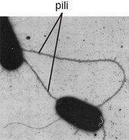
Fimbriae and pili are proteinaceous appendages on prokaryotic cells.
Fimbriae: Short, numerous, used for attachment to surfaces and biofilm formation.
Pili: Longer, fewer, involved in attachment to other cells and DNA transfer (conjugation).
Bacterial Morphologies and Arrangements
Bacteria exhibit a limited number of shapes and arrangements, which are important for identification.
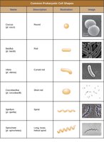
Common Shapes: Coccus (round), Bacillus (rod), Spirillum (spiral), Spirochete (flexible spiral).
Arrangements: Single, diplo (pairs), tetrads (fours), staphylo (clusters), strepto (chains).
Bacterial Cell Wall
Structure and Function
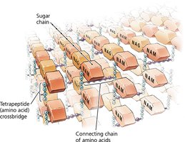
The cell wall is a rigid structure that provides shape, protection from osmotic pressure, and is a key virulence factor. It is primarily composed of peptidoglycan.
Peptidoglycan: A mesh-like polymer of sugars (N-acetylglucosamine and N-acetylmuramic acid) and amino acids.
Gram-Positive vs. Gram-Negative Cell Walls
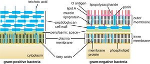
Bacteria are classified based on their cell wall structure, which affects their staining properties and susceptibility to antibiotics.
Gram-Positive: Thick peptidoglycan layer, contains teichoic acids.
Gram-Negative: Thin peptidoglycan layer, outer membrane with lipopolysaccharide (LPS), contains porins.
Plasma Membrane
Structure and Function
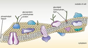
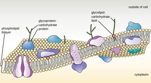
The plasma membrane is a selectively permeable barrier composed of a phospholipid bilayer with embedded proteins. It regulates the movement of substances into and out of the cell.
Phospholipids: Amphipathic molecules with hydrophilic heads and hydrophobic tails.
Fluid Mosaic Model: Describes the dynamic and flexible nature of the membrane.
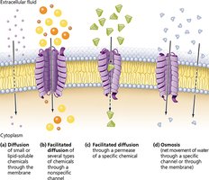
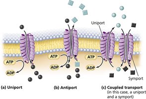
Transport Mechanisms
Passive Transport: Movement down a concentration gradient; includes simple diffusion, facilitated diffusion, and osmosis. No energy required.
Active Transport: Movement against a concentration gradient; requires energy (ATP) and transport proteins.
Osmosis and Tonicity
Osmosis is the diffusion of water across a selectively permeable membrane. The tonicity of the surrounding solution affects cell volume:
Hypertonic: Water leaves the cell, causing plasmolysis (cell shrinks).
Isotonic: No net water movement; cell remains unchanged.
Hypotonic: Water enters the cell, causing it to swell.
Internal Structures of Prokaryotic Cells
Cytoplasm and Cytosol
The cytoplasm is the site of most cellular activities and contains the cytosol, a gel-like matrix composed of water, proteins, carbohydrates, lipids, and ions.
DNA, Ribosomes, and Plasmids
Nucleoid: Region containing the bacterial chromosome (circular DNA).
Plasmids: Small, extrachromosomal DNA molecules carrying non-essential but advantageous genes.
Ribosomes: Sites of protein synthesis; prokaryotic ribosomes are 70S (composed of 50S and 30S subunits).
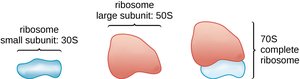
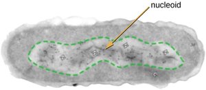
Inclusions and Cytoskeleton
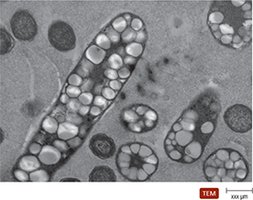
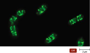
Inclusions are storage granules for nutrients and other substances. The cytoskeleton provides structural support and shape to the cell.
Endospores
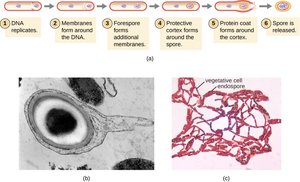
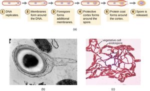
Endospores are highly resistant, dormant structures formed by certain bacteria (e.g., Bacillus and Clostridium) for survival under adverse conditions.
Properties: Resistant to heat, drying, chemicals, and radiation.
Sporulation: The process of endospore formation.
Eukaryotic Cell Structure
General Features
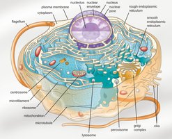
Eukaryotic cells are more complex, containing a true nucleus and various membrane-bound organelles, each with specialized functions.
Eukaryotic Cell Wall
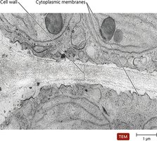
Some eukaryotes have cell walls composed of polysaccharides:
Plants: Cellulose
Fungi: Cellulose, chitin, glucomannan
Algae: Cellulose, agar, carrageenan, silicates, algin, calcium carbonate
Plasma Membrane
The eukaryotic plasma membrane is similar to that of prokaryotes, consisting of a phospholipid bilayer with embedded proteins and carbohydrates.
Motility: Flagella and Cilia
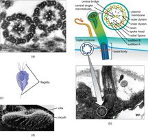
Eukaryotic cells may possess flagella or cilia for movement. Eukaryotic flagella undulate, while cilia move with a rowing motion.
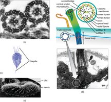
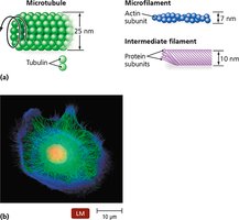
Cytoskeleton
The cytoskeleton is an extensive network of protein fibers that maintains cell shape, anchors organelles, and facilitates movement.
Endomembrane System
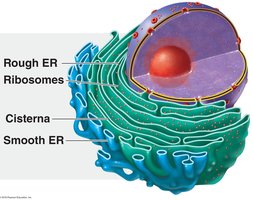
The endomembrane system is unique to eukaryotes and is responsible for synthesizing and transporting cellular components. It includes the endoplasmic reticulum (ER), Golgi apparatus, lysosomes, and vesicles.
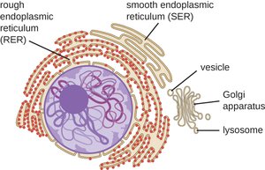
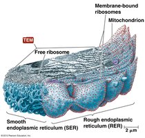
Endoplasmic Reticulum (ER)
Rough ER: Studded with ribosomes; site of protein synthesis.
Smooth ER: Lacks ribosomes; synthesizes lipids and carbohydrates.
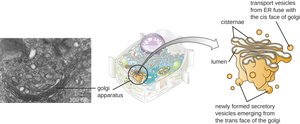
Golgi Complex
The Golgi complex processes, sorts, and ships proteins and lipids produced by the ER. It packages molecules into vesicles for transport.
Lysosomes, Peroxisomes, and Vacuoles
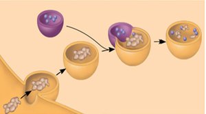
Lysosomes: Contain hydrolytic enzymes for digestion; function at acidic pH.
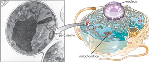
Peroxisomes: Contain oxidase and catalase; degrade toxic substances.
Vacuoles: Temporary storage compartments.
Mitochondria and Chloroplasts
Mitochondria: Site of ATP production via cellular respiration; contain their own DNA and ribosomes.
Chloroplasts: Found in photosynthetic organisms; site of photosynthesis; contain thylakoids, DNA, and ribosomes.
Nucleus
The nucleus contains most of the cell's genetic material, is surrounded by a nuclear envelope, and contains nuclear pores for molecular exchange. The nucleolus is the site of rRNA synthesis.
Summary Comparison: Prokaryotic vs. Eukaryotic Cells
Both: Have plasma membranes, cytoplasm, ribosomes, and genetic material.
Differences: Eukaryotes have a nucleus and membrane-bound organelles; prokaryotes do not.
Table: Comparison of Prokaryotic and Eukaryotic Cell Features
Feature | Prokaryotic Cells | Eukaryotic Cells |
|---|---|---|
Nucleus | Absent | Present |
Membrane-bound organelles | Absent | Present |
Cell wall | Usually present (peptidoglycan) | Present in some (cellulose, chitin, etc.) |
Size | ~1 µm or less | 10–100 µm |
Ribosomes | 70S | 80S (cytoplasm), 70S (organelles) |
Example: Escherichia coli is a prokaryote; human cells are eukaryotic.
