 Back
BackChapter 15: Diagnosing Infections – Microbial Identification and Laboratory Techniques
Study Guide - Smart Notes
 Tailored notes based on your materials, expanded with key definitions, examples, and context.
Diagnosing Infections: Overview
Diagnosing infectious diseases is a critical aspect of clinical microbiology, involving the identification of pathogens responsible for disease. Accurate diagnosis guides effective treatment and infection control. This chapter reviews the main categories of microbial identification, specimen collection, and the laboratory techniques used in clinical microbiology.
Major Categories of Microbial Identification Techniques
Overview of Identification Methods
Phenotypic Methods: Involve the observation of observable traits such as morphology, physiology, and biochemical properties.
Immunologic Methods: Utilize serological analysis to detect specific antibodies or antigens in patient samples.
Genotypic Methods: Analyze the genetic material (DNA or RNA) of the microorganism for identification.
Combining data from these methods provides a unique profile for any bacterium, enhancing diagnostic accuracy.
Phenotypic Methods
Principles and Applications
Phenotypic methods focus on the traits that an organism expresses, including its appearance and behavior. These methods are foundational in microbiology laboratories.
Microscopic and Macroscopic Morphology: Examining cell shape, arrangement, and staining characteristics.
Physiological and Biochemical Testing: Assessing metabolic capabilities, such as enzyme activities and fermentation patterns.
Antibiotic Susceptibility: Determining which antibiotics inhibit or kill the organism.
Chemical Composition: Analyzing cell wall and membrane components.
Direct Examination of Specimens
Direct microscopic observation of fresh or stained specimens is a rapid method for presumptive identification.
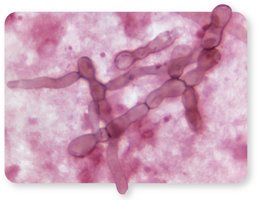
Common Stains: Gram stain, acid-fast stain (for Mycobacterium), and KOH preparation (for fungi).
Growth-Based Methods
Some phenotypic methods require culturing the organism on specialized media:
Selective Media: Encourage the growth of suspected pathogens while inhibiting others.
Differential Media: Distinguish organisms based on metabolic reactions, such as fermentation.
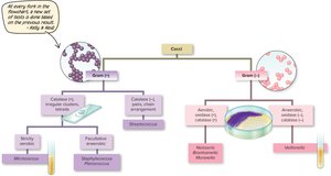
Dichotomous Key
A dichotomous key is a flowchart that guides identification based on sequential test results, such as Gram stain and enzymatic tests.
Biochemical Testing
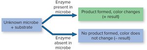
Biochemical tests detect enzyme-mediated metabolic reactions, often visualized by color changes in the medium.
Principle: The microbe is cultured with a substrate; if the enzyme is present, a product forms and the color changes.
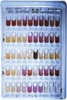
Example: API test strips for rapid identification of bacteria based on multiple biochemical reactions.
Drawbacks of Phenotypic Methods
Culture-based methods are time-consuming (minimum 18–24 hours).
Some pathogens are nonculturable, leading to possible misidentification.
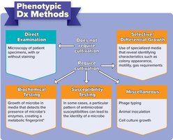
Summary Table: Phenotypic Diagnostic Methods
Method | Description |
|---|---|
Direct Examination | Microscopy of patient specimens, with or without staining |
Selective/Differential Growth | Use of specialized media to reveal identifying characteristics |
Biochemical Testing | Growth in media detecting metabolic activities |
Susceptibility Testing | Pattern of antimicrobial susceptibility |
Miscellaneous | Phage typing, animal inoculation, cell culture growth |
Immunologic Methods
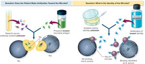
Serological Testing Principles
Immunologic methods detect the presence of specific antibodies or antigens in patient samples, exploiting the specificity of the immune response.
Applications: Determining immunologic status, confirming diagnoses, and screening for diseases.
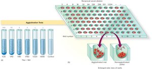
Agglutination and Precipitation Reactions
These reactions involve the visible clumping or precipitation of antigens and antibodies.
Agglutination: Whole cells or organisms are cross-linked by antibodies, forming visible clumps.
Precipitation: Soluble antigens form insoluble complexes with antibodies, which may be harder to visualize.
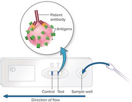
Immunochromatography (Lateral Flow Tests)
These rapid tests use a cartridge with antibodies to detect antigens in patient samples, producing a visible color change if positive.
Examples: Pregnancy tests, rapid strep tests.
Antibody Titers
The concentration of antibodies is determined by serial dilution and agglutination testing. The highest dilution that still produces agglutination reflects the antibody titer.
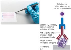
Western Blot Procedure
Western blotting separates microbial proteins by electrophoresis, transfers them to a membrane, and detects specific antibodies or antigens using labeled antibodies. It is used to verify the presence of microbial-specific antigens or antibodies.
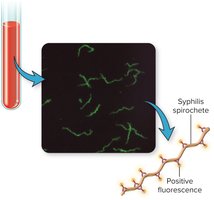
Immunofluorescence Testing
Fluorescently labeled antibodies bind to specific antigens, allowing visualization under a fluorescence microscope. This method is valuable for identifying pathogens in tissues or fluids.
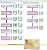
Enzyme-Linked Immunosorbent Assay (ELISA)
ELISA uses enzyme-linked antibodies to detect antigen-antibody reactions, producing a color change. There are two main types:
Indirect ELISA: Detects antibodies in patient serum using a known antigen.
Direct ELISA: Detects antigens using a known antibody.
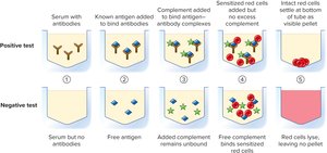
Complement Fixation Test
This test detects the presence of specific antibodies by their ability to fix complement and prevent lysis of red blood cells. It is used for diagnosing viral and fungal diseases.
Genotypic Methods
Polymerase Chain Reaction (PCR)
PCR amplifies specific DNA or RNA sequences, allowing detection of even minute quantities of microbial genetic material. Real-time PCR (qPCR) uses fluorescent labeling for rapid, quantitative results. Multiplex PCR can detect multiple pathogens simultaneously.
Example: RT-PCR for SARS-CoV-2 (COVID-19) diagnosis.
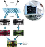
Hybridization and Microarrays
Hybridization uses labeled DNA or RNA probes to detect complementary sequences in microbial nucleic acids. Microarrays ("chips") contain thousands of gene sequences, allowing simultaneous detection of multiple pathogens.
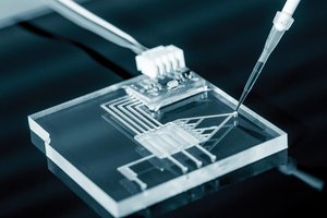
Lab on a Chip
Miniaturized genetic tests on integrated circuits (chips) enable rapid, point-of-care diagnostics with minimal supplies and training. These are especially valuable in resource-limited settings.
Specimen Collection and Handling
Importance of Proper Collection
Accurate identification depends on proper specimen collection, handling, storage, and transport. Aseptic technique and sterile containers are essential to prevent contamination. Only the infected site should be sampled, avoiding normal microbiota.
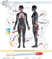
Common Specimen Types
Sputum and Saliva: Sputum is collected by coughing or catheter; avoid saliva contamination.
Urine: Collected aseptically by catheter or "clean catch" method; mucosal samples by swab.
Skin and Wounds: Swabbed, scraped, or biopsied for deeper layers.
Fluids: Blood, cerebrospinal fluid, and tissue fluids collected by sterile needle aspiration.
Other Sites: Eye, ear canal, synovial fluid, nasal cavity, and biopsied tissue.
Laboratory Workflow
Patient analysis includes clinical signs, specimen collection, laboratory testing, and result reporting. Timely and accurate labeling, transport, and patient history are crucial for effective diagnosis.
Summary Table: Comparison of Identification Methods
Method | Principle | Advantages | Limitations |
|---|---|---|---|
Phenotypic | Observation of traits and metabolic activities | Widely available, inexpensive | Time-consuming, some pathogens nonculturable |
Immunologic | Detection of antibodies/antigens | Rapid, specific, can detect past exposure | False positives/negatives possible |
Genotypic | Analysis of genetic material | Highly sensitive, does not require culture | Requires specialized equipment |
Additional info: This chapter aligns with Ch. 15 - Infections and covers laboratory techniques (Ch. 2), microbial growth control (Ch. 9), and diagnostic applications relevant to pathogenesis and epidemiology (Ch. 11).
