Back
BackChapter 16: Adaptive Immunity
Study Guide - Smart Notes
 Tailored notes based on your materials, expanded with key definitions, examples, and context.
Adaptive Immunity: An Overview
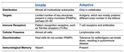
Comparison of Innate and Adaptive Immunity
Adaptive immunity is a specialized branch of the immune system that provides specific, inducible, and long-lasting defense against pathogens. It is distinct from innate immunity in several key aspects:
Innate | Adaptive | |
|---|---|---|
Distribution | Almost all multicellular eukaryotes | Only in vertebrates |
Targets | Limited number of key structures (PAMPs) | Antigens, mostly proteins; highly diverse |
Immune Receptors | Pattern recognition receptors (e.g., TLRs) | T cell receptors and antibodies |
Cellular Presence | Almost all cells | Lymphocytes only |
Discrimination | Host cells lack PAMPs | Tolerance for self-antigens (can break down in autoimmunity) |
Immunological Memory | Absent | Present |
Key Attributes of Adaptive Immunity
Specificity: Targets unique antigens.
Inducibility: Activated in response to specific pathogens.
Clonality: Generates clones of lymphocytes specific to the antigen.
Unresponsiveness to Self: Normally does not attack the body's own cells.
Memory: Remembers previous encounters for faster future responses.
Cells and Organs of Adaptive Immunity
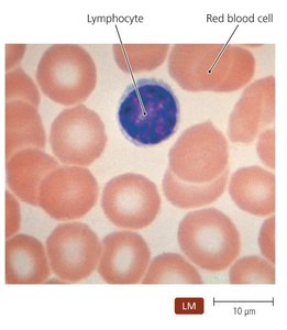
Lymphocytes
Lymphocytes are the central cells of adaptive immunity, including B lymphocytes (B cells) and T lymphocytes (T cells). B cells mature in the bone marrow, while T cells mature in the thymus. Both circulate in the blood and reside in lymphoid organs.
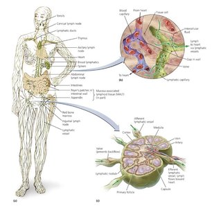
Lymphatic System
The lymphatic system is composed of lymphatic vessels, lymphoid cells, tissues, and organs. It screens the body for foreign molecules and returns lymph to the circulatory system. Primary lymphoid organs include the red bone marrow and thymus; secondary organs include lymph nodes, spleen, tonsils, and MALT (mucosa-associated lymphoid tissue).
Antigens and Epitopes
Definition and Properties
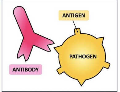
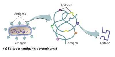
Antigens are molecules recognized as foreign and capable of provoking an immune response. The specific regions recognized by immune receptors are called epitopes or antigenic determinants. Large, complex molecules such as proteins make the best antigens.
Types of Antigens
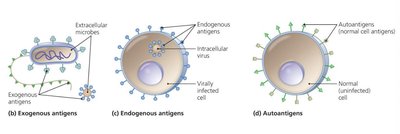
Exogenous antigens: Toxins and components of microbial cell walls, membranes, flagella, and pili.
Endogenous antigens: Produced by microbes that reproduce inside body cells (e.g., viruses).
Autoantigens: Derived from normal cellular processes; can be involved in autoimmune diseases.
Major Histocompatibility Complex (MHC) and Antigen Presentation
MHC Molecules
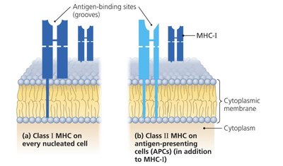
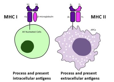
MHC molecules are glycoproteins found on cell membranes that present antigenic peptides to T cells. There are two main classes:
MHC Class I: Present on all nucleated cells; present endogenous antigens.
MHC Class II: Present on antigen-presenting cells (APCs) such as macrophages, B cells, and dendritic cells; present exogenous antigens.
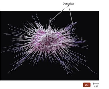
Antigen-Presenting Cells (APCs)
Dendritic cells are the most common APCs, capturing antigens and presenting them to T cells to initiate adaptive responses.
T Lymphocytes (T Cells)
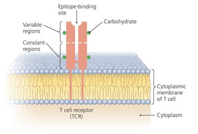
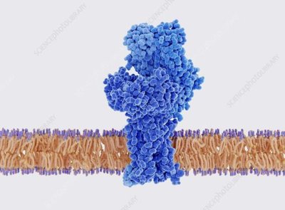
T Cell Receptors (TCRs)
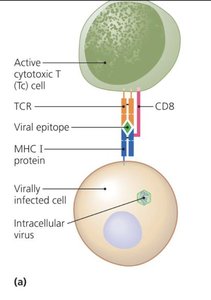
T cells possess T cell receptors (TCRs) that recognize antigenic peptides only when presented by MHC molecules. TCRs do not bind free antigens directly.
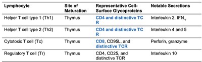
Types of T Lymphocytes
Cytotoxic T lymphocytes (Tc): Directly kill infected or abnormal cells.
Helper T lymphocytes (Th): Regulate B cells and Tc cells; include Th1 and Th2 subtypes.
Regulatory T lymphocytes (Treg): Suppress immune responses to prevent autoimmunity.
Lymphocyte | Site of Maturation | Representative Cell-Surface Glycoproteins | Notable Secretions |
|---|---|---|---|
Helper T cell type 1 (Th1) | Thymus | CD4 and distinctive TCR | Interleukin 2, IFN-γ |
Helper T cell type 2 (Th2) | Thymus | CD4 and distinctive TCR | Interleukin 4 and 5 |
Cytotoxic T cell (Tc) | Thymus | CD8, CD95L, and distinctive TCR | Perforin, granzyme |
Regulatory T cell (Tr) | Thymus | CD4, CD25, and distinctive TCR | Interleukin 10 |
Clonal Deletion of T Cells
Self-reactive T cells are eliminated in the thymus through apoptosis to prevent autoimmune responses.
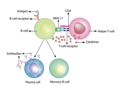
B Lymphocytes (B Cells) and Antibodies
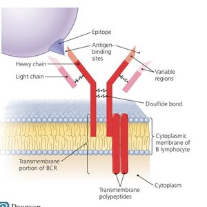
B Cell Receptors (BCRs)
B cells possess B cell receptors (BCRs) that bind specific epitopes directly. Each B cell expresses a unique BCR, allowing the immune system to recognize a vast array of antigens.
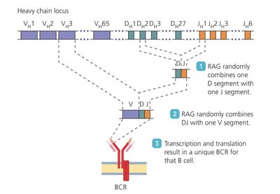
Generation of BCR Diversity
The enzyme RAG recombines variable (V), diversity (D), and joining (J) gene segments to generate diverse BCRs, enabling recognition of millions of different epitopes.
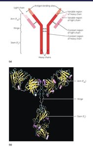
Antibody Structure and Classes
Antibodies (immunoglobulins) are secreted by plasma cells and have antigen-binding sites identical to the BCR of the activated B cell. There are five main classes of antibodies, each with distinct functions and structures:
Class | Structure | Main Functions | Location |
|---|---|---|---|
IgM | Pentamer | First produced, agglutination, complement activation | Serum |
IgG | Monomer | Most abundant, opsonization, neutralization, crosses placenta | Serum |
IgA | Dimer (secretory) | Secretions, mucosal immunity | Secretions, serum |
IgE | Monomer | Allergy, antiparasitic responses | Serum, bound to mast cells |
IgD | Monomer | Unclear function, B cell receptor | B cell surface |
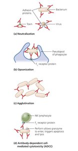
Antibody Functions
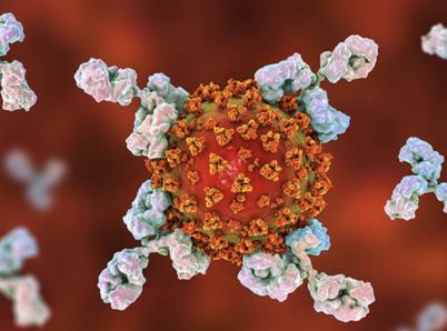
Neutralization: Blocks pathogen attachment or toxin activity.
Opsonization: Enhances phagocytosis by marking pathogens.
Agglutination: Clumps pathogens for easier clearance.
Complement Activation: Triggers lysis and inflammation.
Antibody-Dependent Cellular Cytotoxicity (ADCC): Directs killing by immune cells.
Clonal Deletion of B Cells
Self-reactive B cells are eliminated or inactivated in the bone marrow to prevent autoimmunity.
Cytokines in Immune Regulation
Definition and Types
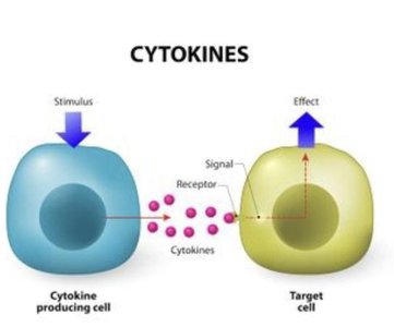
Cytokines are soluble proteins that mediate communication between immune cells. Major types include:
Interleukins (ILs): Signal among leukocytes.
Interferons (IFNs): Antiviral and immune-modulating proteins.
Growth Factors: Stimulate cell division.
Tumor Necrosis Factor (TNF): Inflammation and apoptosis.
Chemokines: Attract immune cells to sites of infection.
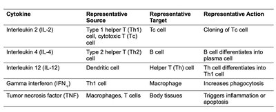
Cytokine | Source | Target | Action |
|---|---|---|---|
IL-2 | Th1, Tc cells | Tc cell | Cloning of Tc cell |
IL-4 | Th2 cell | B cell | B cell differentiation |
IL-12 | Dendritic cell | Th1 cell | Th1 differentiation |
IFN-γ | Th1 cell | Macrophage | Increases phagocytosis |
TNF | Macrophages, T cells | Body tissues | Triggers inflammation/apoptosis |
Cell-Mediated Immune Responses
Activation and Function of Cytotoxic T Cells
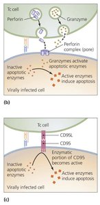
Cytotoxic T cells (Tc) recognize and kill infected or abnormal cells via two main pathways:
Perforin-Granzyme Pathway: Tc cells release perforin (forms pores) and granzymes (induce apoptosis).
CD95 Pathway: Tc cells engage CD95 on target cells, triggering apoptosis.
Memory T Cells
Some activated T cells become memory T cells, which persist long-term and respond rapidly upon re-exposure to the same antigen.
Antibody (Humoral) Immune Responses
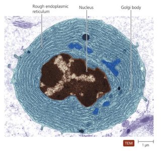
Plasma Cells and Antibody Production
Plasma cells are differentiated B cells that secrete large amounts of antibodies specific to the encountered antigen. These cells are short-lived, but the antibodies and memory B cells they produce provide lasting immunity.
Immunological Memory
Memory B cells persist in lymphoid tissues and enable a rapid, robust secondary immune response upon re-exposure to the same antigen. The primary response is slower and produces fewer antibodies, while the secondary response is faster and stronger.
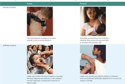
Types of Acquired Immunity
Active vs. Passive, Natural vs. Artificial
Naturally Acquired Active Immunity: Immune response to antigens encountered in daily life (e.g., infection).
Naturally Acquired Passive Immunity: Transfer of antibodies from mother to child (e.g., via placenta or breast milk).
Artificially Acquired Active Immunity: Immune response to antigens introduced by vaccination.
Artificially Acquired Passive Immunity: Transfer of antibodies (e.g., antitoxins) by injection.