 Back
BackChapter 3: Bacteria and Archaea – Structure, Function, and Classification
Study Guide - Smart Notes
 Tailored notes based on your materials, expanded with key definitions, examples, and context.
Bacteria and Archaea: An Overview
Prokaryotic Cell Structure and Distinctions
Bacteria and archaea are prokaryotic microorganisms that differ fundamentally from eukaryotes in several ways:
DNA Packaging: Prokaryotes lack a nucleus and histones, resulting in a different organization of genetic material.
Cell Wall Composition: Bacterial cell walls contain peptidoglycan, while archaea have unique chemical structures.
Internal Structures: Prokaryotes lack membrane-bound organelles found in eukaryotes.
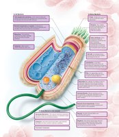
The Structure of the Bacterial Cell
Essential and Variable Structures
All bacterial cells possess certain fundamental structures, while others are present only in some species.
All bacteria have:
Cytoplasmic membrane
Cytoplasm
Ribosomes
Cytoskeleton
One (or a few) chromosome(s)
Most bacteria have:
Cell wall
Glycocalyx (surface coating)
Some bacteria have:
Flagella, pili, fimbriae
Outer membrane
Nanowires/nanotubes
Plasmids
Inclusions
Endospores
Microcompartments
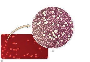
Bacterial Shapes and Arrangements
Major Shapes of Bacteria
Bacteria exhibit a variety of shapes and arrangements, which are important for identification and classification.
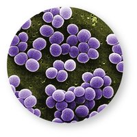
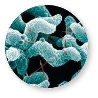
Coccus (plural: cocci): Spherical or ball-shaped cells. Can be oval, bean-shaped, or pointed.
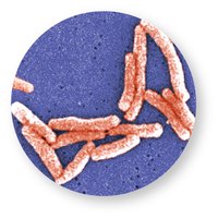
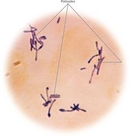
Bacillus (plural: bacilli): Cylindrical or rod-shaped cells. Variations include blocky, spindle-shaped, filamentous, club-shaped, or drumstick-shaped. Short rods are called coccobacilli.
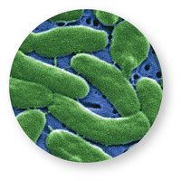
Vibrio: Gently curved rods.
Spirillum: Rigid, spiral-shaped cells, twisted like a corkscrew.


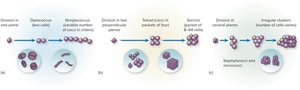
Bacterial Arrangements
Bacterial cells can be found as single cells or in characteristic groupings due to their patterns of division.
Cocci arrangements:
Single
Diplococci (pairs)
Tetrads (groups of four)
Staphylococci/micrococci (irregular clusters)
Streptococci (chains)
Sarcina (cubical packets of 8, 16, or more)
Bacilli arrangements:
Single
Diplobacilli (pairs)
Streptobacilli (chains)
Palisades (partially attached chains)
External Structures of Bacteria
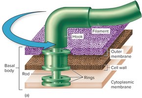
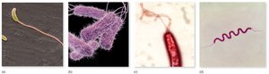
Flagella and Motility
Flagella are appendages used for motility, allowing bacteria to move toward or away from stimuli (chemotaxis).
Structure: Composed of filament, hook, and basal body.
Arrangements:
Monotrichous: single flagellum
Lophotrichous: small bunches at one end
Amphitrichous: flagella at both ends
Peritrichous: flagella all over the cell surface
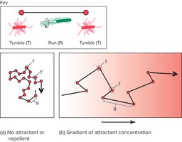
Function: Movement is achieved by rotation; counterclockwise rotation results in a run (straight movement), while clockwise rotation causes a tumble (change in direction).
Chemotaxis
Bacteria move in response to chemical gradients:
Positive chemotaxis: Movement toward attractants (e.g., nutrients).
Negative chemotaxis: Movement away from repellents.
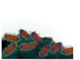
Other Appendages
Fimbriae: Small, bristle-like fibers for adhesion to surfaces and host tissues.
Pili: Tubular structures used for conjugation (DNA transfer) and sometimes motility.
Nanotubes/Nanowires: Tubular extensions for transferring nutrients or electrons.
Surface Layers: S Layer and Glycocalyx
S Layer
A single layer of protein subunits, produced in hostile environments for protection or attachment.
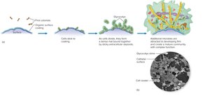
Glycocalyx
A coating of polysaccharide or glycoprotein units outside the cell wall. It can be a loose slime layer or a dense capsule.
Capsule: Tightly bound, protects against phagocytosis, and enhances pathogenicity.
Biofilm formation: Glycocalyx enables bacteria to adhere to surfaces and form persistent communities (biofilms).
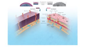
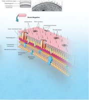
The Bacterial Cell Envelope
Gram-Positive vs. Gram-Negative Cell Walls
The cell envelope consists of layers that protect and support the cell. The structure differs between Gram-positive and Gram-negative bacteria:
Gram-Positive: Thick peptidoglycan layer, teichoic acids, no outer membrane.
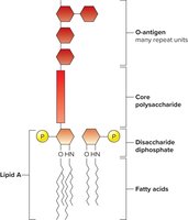
Gram-Negative: Thin peptidoglycan layer, outer membrane with lipopolysaccharide (LPS), periplasmic space.
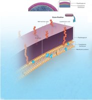
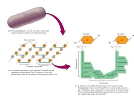
Peptidoglycan Structure
Peptidoglycan is a mesh-like polymer of sugars and amino acids, providing rigidity and shape to the cell wall.
Alternating sugars: N-acetylglucosamine (G) and N-acetylmuramic acid (M)
Cross-linked by short peptide chains
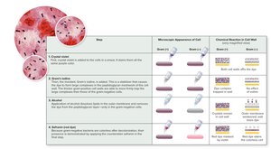
Gram Stain Procedure
The Gram stain differentiates bacteria based on cell wall structure:
Crystal violet (primary stain)
Gram's iodine (mordant)
Alcohol (decolorizer)
Safranin (counterstain)
Internal Structures of Bacteria
Cytoplasm
The cytoplasm is a water-based solution containing sugars, amino acids, salts, and other molecules necessary for cell function.
Genetic Material
Bacterial chromosome: Single, circular DNA molecule located in the nucleoid region.
Plasmids: Small, circular DNA molecules carrying nonessential but advantageous genes (e.g., antibiotic resistance).
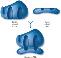
Ribosomes
Sites of protein synthesis, composed of rRNA and protein. Bacterial ribosomes are 70S (30S small subunit + 50S large subunit).
Inclusion Bodies and Microcompartments
Storage sites for nutrients, gases, or magnetic particles. Microcompartments are protein-coated packets containing enzymes for specific pathways.
Cytoskeleton
Protein filaments arranged in helical ribbons, contributing to cell shape and division. Chemically distinct from eukaryotic cytoskeletons.
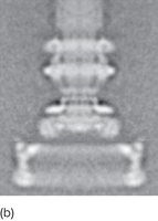
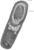
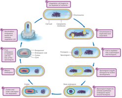
Endospores
Dormant, highly resistant structures formed by genera such as Bacillus and Clostridium. Endospores enable survival in extreme conditions.
Archaea: Unique Features
Distinctive Characteristics
Archaea are a separate domain of prokaryotes, more closely related to eukaryotes than bacteria in some aspects:
Unique rRNA sequences
Distinct membrane lipids and cell wall components
Extremophiles: thrive in extreme environments (temperature, salinity, acidity)
Classification of Bacteria and Archaea
Bergey’s Manuals
Bergey’s Manual of Systematic Bacteriology: Based on genetic relatedness (rRNA sequencing).
Bergey’s Manual of Determinative Bacteriology: Based on phenotypic characteristics (shape, metabolism).
Major Divisions Based on Cell Wall Structure
Division | Characteristics |
|---|---|
Gracilicutes | Gram-negative, thin cell walls |
Firmicutes | Gram-positive, thick, strong cell walls |
Tenericutes | Lack a cell wall, soft |
Mendosicutes | Archaea, unusual cell walls and nutritional habits |
Species and Subspecies
Species: Collection of cells sharing similar traits (≥95% gene similarity).
Subspecies/Strain/Type: Variants within a species with distinct characteristics.
Serotype: Subspecies that elicit unique antibody responses due to surface molecules.
