Back
BackDNA Viruses: Structure, Classification, and Human Diseases
Study Guide - Smart Notes
 Tailored notes based on your materials, expanded with key definitions, examples, and context.
DNA Viruses: Classification and General Features
Overview of DNA Virus Families
DNA viruses causing human disease are classified into seven families based on their DNA type, envelope presence, size, and host specificity.
dsDNA Viruses: Poxviridae, Herpesviridae, Papillomaviridae, Polyomaviridae, Adenoviridae
ssDNA Viruses: Parvoviridae
Mixed dsDNA/ssDNA: Hepadnaviridae
Envelope: Some families possess an envelope, affecting transmission and immune evasion.
Host Range: Viruses may be species-specific or capable of zoonotic transmission.
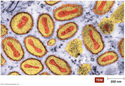
Poxviridae
Structure and Transmission
Genome: Double-stranded DNA
Capsid: Complex structure with envelope
Size: Second largest viruses infecting mammals
Transmission: Primarily via inhalation; close contact required
Diseases Caused by Poxviridae
Smallpox (Variola): Two forms: Variola major and minor. Virus spreads via blood to skin, causing pox and scarring, especially on the face. First human disease eradicated.
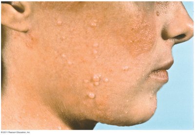
Molluscum Contagiosum: Caused by Molluscipoxvirus. Characterized by smooth, waxy papules on face, trunk, and genitalia. Heals without treatment in immunocompetent individuals.
Animal Poxviruses: Usually species-specific; zoonotic transmission is rare and typically mild.
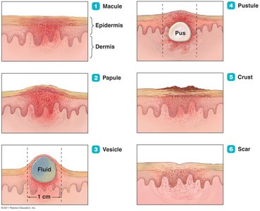
Stages of Poxvirus Lesions
The progression of skin lesions in poxvirus infections follows distinct stages:
Macule: Flat, reddened area
Papule: Raised, solid lesion
Vesicle: Fluid-filled blister
Pustule: Pus-filled lesion
Crust: Dried exudate
Scar: Permanent mark after healing
Smallpox Lesions
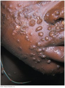
Molluscum Contagiosum Lesions
Herpesviridae
Structure and Latency
Genome: Linear dsDNA
Capsid: Enveloped polyhedral
Entry: Envelope fusion with host cell membrane
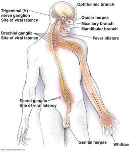
Latency: Virus remains inactive in cells; reactivation causes recurrent disease
Nomenclature: Human herpesviruses (HHV) numbered by discovery order
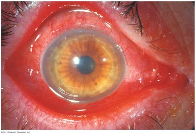
HHV-1 and HHV-2 (Herpes Simplex Viruses)
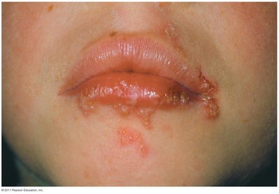
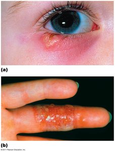
Manifestations: Oral herpes, genital herpes, ocular herpes, whitlow, neonatal herpes
Transmission: Close contact, entry via mucous membrane breaks
Pathogenesis: Virus spreads cell-to-cell via syncytia formation
Epidemiology: HHV-1: casual contact in children; HHV-2: sexual activity (ages 15–29)
Oral Herpes Lesions
Ocular Herpes and Whitlow
Diagnosis, Treatment, and Prevention
Diagnosis: Characteristic lesions
Treatment: Chemotherapeutic agents limit lesion duration and viral shedding; do not cure or eliminate latent virus
Prevention: Gloves for healthcare workers; sexual abstinence or monogamy
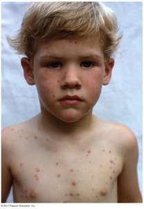
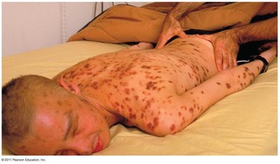
HHV-3 (Varicella-Zoster Virus)
Diseases: Varicella (chickenpox) in children; herpes zoster (shingles) in adults
Transmission: Highly infectious; entry via respiratory tract or eyes
Pathogenesis: Virus travels via blood; skin lesions appear 2–3 weeks post-infection
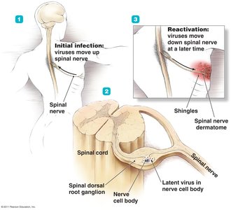
Latency: Virus can reactivate as shingles
Latency and Reactivation of VZV
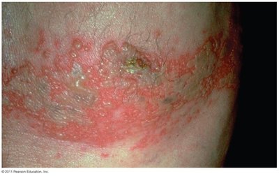
Shingles Lesions
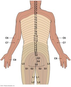
Dermatomes
Diagnosis, Treatment, and Prevention
Diagnosis: Chickenpox diagnosed by lesions; shingles more difficult
Treatment: Chickenpox is self-limiting; shingles managed symptomatically
Prevention: Difficult due to viral shedding before symptoms
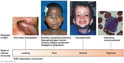
HHV-4 (Epstein-Barr Virus)
Diseases: Infectious mononucleosis, Burkitt’s lymphoma, nasopharyngeal cancer, oral hairy leukoplakia
Transmission: Saliva
Pathogenesis: Infects pharynx, parotid glands, then B lymphocytes; latent infection suppresses apoptosis
Immune Response: Cytotoxic T cells kill infected B cells; symptoms depend on immune status
Diagnosis, Treatment, and Prevention
Diagnosis: Based on characteristic signs
Treatment: Chemotherapy for Burkitt’s lymphoma; symptomatic relief for mono
Prevention: Difficult due to widespread transmission
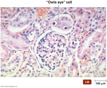
HHV-5 (Cytomegalovirus)
Pathology: Infected cells become enlarged (cytomegaly)
Transmission: Bodily secretions; requires close contact and large exchange
Complications: Severe in fetuses, newborns, immunodeficient patients
Diagnosis, Treatment, and Prevention
Diagnosis: Detection of enlarged cells and cellular inclusions
Treatment: Difficult in fetuses/newborns; Fomiversen for CMV eye infections
Prevention: Abstinence and safe sex
Other Herpesvirus Infections
HHV-6 (Roseolovirus): Causes roseola; pink rash, possible link to multiple sclerosis
HHV-8 (Rhadinovirus): Associated with Kaposi’s sarcoma
Papillomaviridae and Polyomaviridae
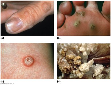
Papillomavirus Infections
Diseases: Papillomas (warts) on skin/mucous membranes; genital warts linked to cancer
Transmission: Direct contact, fomites, autoinoculation
Diagnosis: Observation of papillomas; PAP smear for genital cancers
Treatment: Removal of warts; prevention difficult except for genital warts (abstinence/monogamy)
Polyomavirus Infections
Diseases: Tumors, urinary tract infections (BK virus), progressive multifocal leukoencephalopathy (JC virus)
Pathogenesis: Latent infections in kidneys; reactivation depends on immune status
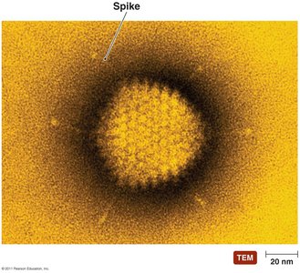
Adenoviridae
Structure and Diseases
Genome: Single, linear dsDNA
Diseases: Common cold, mild diarrhea, conjunctivitis (pinkeye)
Transmission: Respiratory droplets, endocytosis in respiratory tract
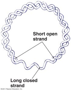
Hepadnaviridae
Structure and Replication
Genome: Both single- and double-stranded DNA
Replication: Via RNA intermediary (unique among DNA viruses)
Includes: Hepatitis B virus (HBV)
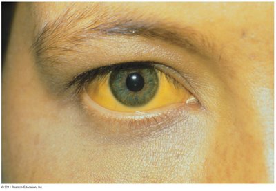
Hepatitis B Infections
Disease: Hepatitis (liver inflammation), jaundice, liver enlargement, abdominal distress, bleeding
Transmission: Saliva, semen, vaginal secretions; needles, sexual intercourse, childbirth
Complications: Coinfection with hepatitis D increases risk of permanent liver damage; associated with liver cancer
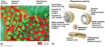
Viral Particles and Diagnosis
Body Fluids: Contain three types of virus particles
Diagnosis: Detection of viral antigens
Treatment: No universally effective treatment; prevention via vaccination and safer sexual practices
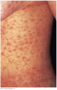
Parvoviridae
Structure and Disease
Genome: ssDNA (only human pathogen with this genome)
Size: Smallest DNA virus
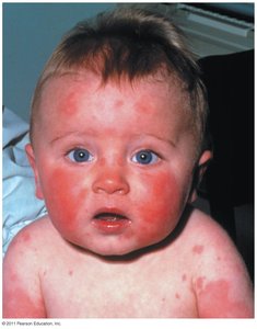
Disease: B19 virus causes erythema infectiosum (fifth disease); characterized by skin reddening, aggravated by sunlight