 Back
BackEukaryotic Cell Structure and Function: Origins, Anatomy, and Membrane Transport
Study Guide - Smart Notes
 Tailored notes based on your materials, expanded with key definitions, examples, and context.
Eukaryotic Cell Structure and Function
Origin of Eukaryotic Cells
The origin of eukaryotic cells is a foundational concept in microbiology, explaining the evolutionary steps that led to complex cellular life. Eukaryotes are distinguished from prokaryotes by their compartmentalized structures and organelles.
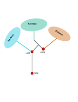
First Universal Common Ancestor (FUCA): The earliest ancestor, likely non-cellular, composed of RNA and proteins.
Last Universal Common Ancestor (LUCA): The ancestor from which all modern life (bacteria, archaea, eukaryotes) evolved.
Last Eukaryotic Common Ancestor (LECA): The ancestor of all eukaryotes, existing approximately 1.8–2.4 billion years ago.
Endosymbiotic Theory
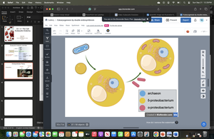
The endosymbiotic theory is the most widely accepted explanation for the origin of mitochondria and chloroplasts in eukaryotic cells. It proposes that a primordial archaeal cell engulfed free-living bacteria, which then became permanent residents, evolving into organelles.
Mitochondrial Endosymbiosis: An aerobic bacterium was engulfed by an archaeal host, providing the host with enhanced energy production (ATP) and oxygen detoxification.
Mutual Benefit: The host cell offered protection, while the bacterium supplied energy.
Evidence for Endosymbiosis: Mitochondria and chloroplasts have their own circular DNA, double membranes, and reproduce independently within the cell, similar to bacteria.
Eukaryotic Cell Anatomy
Overview of Eukaryotic Cell Structure
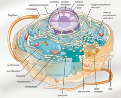
Eukaryotic cells are characterized by membrane-bound organelles, a nucleus, and complex internal structures. They can be unicellular or multicellular and include animals, plants, fungi, and protists.
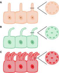
Flagella and Cilia
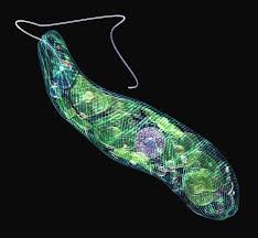
Eukaryotic Flagella
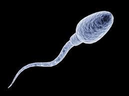
Flagella are whip-like structures used for locomotion in some eukaryotic cells.
Structure: Composed of microtubules in a 9+2 arrangement, surrounded by the plasma membrane, and anchored by a basal body.
Function: Propeller-like motion for movement; sometimes involved in virulence.
Examples: Sperm cells, protozoans such as Euglena.
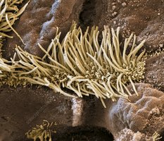
Eukaryotic Cilia
Cilia are shorter and more numerous than flagella, with similar internal structure but distinct functions.
Motile Cilia: Used for locomotion (e.g., in Paramecium) or moving substances across cell surfaces (e.g., in respiratory epithelium).
Non-motile Cilia: Serve as sensory organelles.

Cell Walls in Eukaryotes
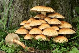
Some eukaryotes possess cell walls, which provide structural support and protection.
Fungi: Cell walls made of chitin (a polysaccharide) and proteins.
Plants: Cell walls composed of cellulose and pectins.

Plasma Membrane Structure and Function
The plasma membrane is a semi-permeable lipid bilayer that regulates the movement of substances into and out of the cell.
Structure: Fluid mosaic of phospholipids, proteins, and cholesterol.
Function: Transport, cell signaling, and maintaining homeostasis.
Transport Mechanisms: Osmosis, simple diffusion, facilitated diffusion, active transport, endocytosis (pinocytosis, phagocytosis, receptor-mediated).
Membrane Transport Mechanisms
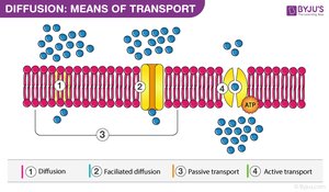
Passive Transport
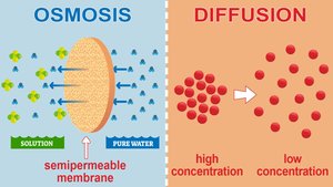
Passive transport moves substances down their concentration gradient without energy input.
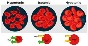
Osmosis: Movement of water across a semi-permeable membrane from high to low water concentration.
Simple Diffusion: Movement of small, non-polar molecules (e.g., O2, CO2).
Facilitated Diffusion: Movement of larger or polar molecules via transport proteins (e.g., glucose, amino acids, ions).
Active Transport
Active transport requires energy (usually ATP) to move substances against their concentration gradient.
Examples: Sodium-potassium pump, uptake of glucose in intestines.

Endocytosis
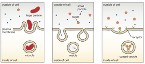
Endocytosis is the process by which cells engulf external substances, forming vesicles.
Phagocytosis: Engulfment of large particles or cells.
Pinocytosis: Uptake of fluids and dissolved substances.
Receptor-mediated Endocytosis: Specific uptake of molecules via receptor-ligand interactions.
Internal Structures and Organelles
Cytoplasm
The cytoplasm is a gel-like matrix that supports organelles, facilitates metabolic reactions, and helps maintain cell shape.
Nucleus and Nuclear Components
The nucleus is the control center of the cell, housing genetic material and coordinating activities such as growth and reproduction.
Nucleus: Contains DNA; site of DNA replication and transcription.
Nucleolus: Site of rRNA synthesis and ribosome assembly.
Nuclear Envelope: Double membrane with pores for molecular transport.

Endoplasmic Reticulum (ER)
Rough ER: Studded with ribosomes; synthesizes and modifies proteins for secretion or membrane insertion.
Smooth ER: Lacks ribosomes; involved in lipid and steroid synthesis, detoxification, and calcium storage.
Golgi Apparatus and Lysosomes
Golgi Apparatus: Modifies, sorts, and packages proteins and lipids for delivery.
Lysosomes: Contain hydrolytic enzymes for digestion of biomolecules.
Ribosomes
Ribosomes are complexes of rRNA and proteins that synthesize proteins from mRNA templates.
Mitochondria
Mitochondria are the powerhouses of the cell, converting glucose into ATP through cellular respiration. They have their own DNA and double membranes, supporting the endosymbiotic theory.
Structure: Inner (cristae) and outer membranes, matrix, circular DNA.
Function: ATP production via aerobic respiration.
Summary Table: Key Eukaryotic Cell Structures
Organelle/Structure | Main Function | Key Features |
|---|---|---|
Nucleus | Genetic information storage, transcription | Double membrane, nuclear pores, nucleolus |
Rough ER | Protein synthesis and modification | Ribosome-studded membranes |
Smooth ER | Lipid synthesis, detoxification | No ribosomes, tubular structure |
Golgi Apparatus | Protein and lipid modification, sorting | Stacked membrane sacs |
Lysosome | Digestion of biomolecules | Enzyme-filled vesicles |
Mitochondria | ATP production | Double membrane, own DNA |
Plasma Membrane | Selective barrier, transport, signaling | Lipid bilayer, proteins, cholesterol |
Cell Wall (plants/fungi) | Structural support | Cellulose (plants), chitin (fungi) |
Flagella/Cilia | Movement, sensory functions | 9+2 microtubule arrangement |
