 Back
BackEukaryotic Cells and Microorganisms: Structure, Function, and Diversity
Study Guide - Smart Notes
 Tailored notes based on your materials, expanded with key definitions, examples, and context.
Eukaryotic Cells and Microorganisms
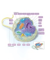
Overview of the Eukaryotic Cell
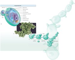
Eukaryotic cells are structurally complex and form the basis of all multicellular life. They evolved from primitive single-celled organisms, which aggregated into colonies and eventually specialized into complex multicellular organisms. Eukaryotic cells contain membrane-bound organelles that compartmentalize cellular functions.
External Structures: Flagella and Cilia
Eukaryotic cells may possess appendages for movement, such as flagella and cilia. These structures are distinct from their prokaryotic counterparts in both size and complexity.
Flagella: Thicker and more complex than bacterial flagella, composed of a sheath and a core of microtubules arranged in a 9 + 2 pattern.
Cilia: Shorter and more numerous than flagella, found in certain protozoa and animal cells, also with a 9 + 2 microtubule arrangement.
Example: Cilia in the human respiratory tract help move mucus and trapped particles out of the lungs.
The Glycocalyx
The glycocalyx is the outermost layer of some eukaryotic cells, composed mainly of polysaccharides. It appears as a network of fibers, a slime layer, or a capsule, and functions in protection, adherence, and signal reception.
Boundary Structures: The Cell Wall
Cell walls provide structural support and shape to certain eukaryotes, such as fungi. Fungal cell walls are chemically distinct from those of bacteria and archaea, consisting of a thick inner layer of chitin or cellulose and a thin outer layer of mixed glycans.
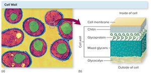
Example: The rigidity of fungal cell walls allows fungi to thrive in diverse environments.
Boundary Structures: The Cell Membrane
The eukaryotic cell membrane is a phospholipid bilayer embedded with proteins and sterols, which provide stability. It acts as a selectively permeable barrier, similar to bacterial and archaeal membranes.
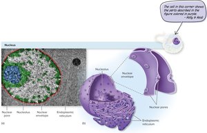
Internal Structures: The Nucleus
The nucleus is the most prominent organelle in eukaryotic cells, containing the cell's genetic material. It is surrounded by a double-membraned nuclear envelope with pores that regulate molecular traffic.
Nucleolus: Site of ribosomal RNA synthesis and ribosome assembly.
Chromatin: Linear DNA complexed with histone proteins.
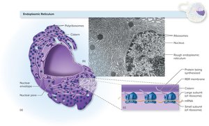
Internal Structures: Endoplasmic Reticulum (ER)
The ER is a network of membranes involved in transport and synthesis. There are two types:
Rough ER (RER): Studded with ribosomes; synthesizes and transports proteins.
Smooth ER (SER): Lacks ribosomes; involved in lipid synthesis and detoxification.
Internal Structures: Golgi Apparatus
The Golgi apparatus modifies, sorts, and packages proteins and lipids for secretion or delivery to other organelles. It consists of flattened sacs called cisternae and works closely with the ER.
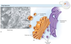
Nature’s Assembly Line: Protein Transport
The nucleus, ER, and Golgi apparatus coordinate the synthesis, modification, and transport of proteins. Proteins are synthesized in the RER, modified in the Golgi, and transported in vesicles to their destinations.
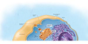
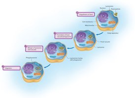
Vesicles: Lysosomes and Vacuoles
Vesicles are membrane-bound sacs involved in storage and transport. Lysosomes contain digestive enzymes for intracellular digestion and defense, while vacuoles store nutrients or waste products.
Mitochondria
Mitochondria are the energy-generating organelles of the cell, extracting energy from nutrients and storing it as ATP. They have a double membrane, their own circular DNA, and 70S ribosomes, supporting the endosymbiotic theory of their origin.
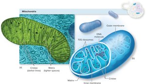
Equation: Aerobic respiration in mitochondria:
Chloroplasts
Chloroplasts are found in algae and plant cells and are responsible for photosynthesis, converting solar energy into chemical energy and producing oxygen as a by-product. They are larger than mitochondria and contain pigments such as chlorophyll.
Ribosomes
Ribosomes are the sites of protein synthesis. Eukaryotic ribosomes are 80S (composed of 60S and 40S subunits), while prokaryotic ribosomes are 70S. Ribosomes may be free in the cytoplasm or attached to the RER.
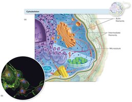
The Cytoskeleton
The cytoskeleton provides structural support, facilitates movement, and organizes cellular components. It consists of three main types of fibers:
Actin filaments: Thin, flexible fibers involved in cell movement and shape changes.
Intermediate filaments: Provide mechanical support.
Microtubules: Hollow tubes that maintain cell shape and serve as tracks for organelle movement.
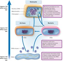
Endosymbiosis
The endosymbiotic theory explains the origin of mitochondria and chloroplasts as descendants of free-living bacteria that were engulfed by ancestral eukaryotic cells. Evidence includes their double membranes, circular DNA, and prokaryote-like ribosomes.
Fungi: Structure, Nutrition, and Reproduction
Fungal Cell Forms and Anatomy
Fungi exist as yeasts (unicellular, round/oval, reproduce by budding) or hyphae (multicellular, threadlike filaments). Some fungi are dimorphic, switching forms based on environmental conditions.
Microscopic Morphology of Yeasts
Yeasts reproduce asexually by budding, forming chains called pseudohyphae. They have a soft, uniform colony texture, while filamentous fungi form cottony or velvety colonies.
Fungal Nutrition
All fungi are heterotrophs, obtaining nutrients from organic substrates. They may be:
Saprobes: Decompose dead organic matter.
Parasites: Infect living hosts, though most do not require a living host.
Fungi secrete enzymes to digest substrates externally and absorb the resulting small molecules.
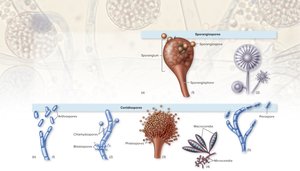
Fungal Reproduction and Spores
Fungi reproduce by forming spores, which can be asexual (sporangiospores, conidiospores) or sexual. Spores are dispersed by air, water, or living things and germinate under favorable conditions.
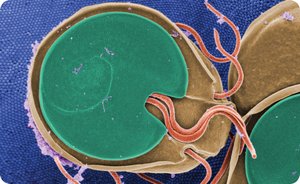
Protozoa: Structure, Life Cycle, and Pathogenicity
Protozoan Form and Function
Protozoa are single-celled eukaryotes with all major organelles. Their cytoplasm is divided into ectoplasm (outer, for movement and feeding) and endoplasm (inner, housing organelles). They move using pseudopods, cilia, or flagella.
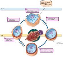
Life Cycle: Trophozoite and Cyst
Protozoa alternate between an active, feeding trophozoite stage and a dormant, resistant cyst stage. The cyst stage allows survival in harsh conditions and facilitates transmission.
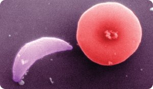
Major Pathogenic Protozoa
Protozoa are classified by their mode of locomotion:
Amoeboid (Sarcodina): Move by pseudopods (e.g., Entamoeba histolytica).
Ciliated (Ciliophora): Move by cilia (e.g., Balantidium coli).
Flagellated (Mastigophora): Move by flagella (e.g., Giardia lamblia).
Apicomplexan (Sporozoa): Nonmotile, complex life cycles (e.g., Plasmodium).
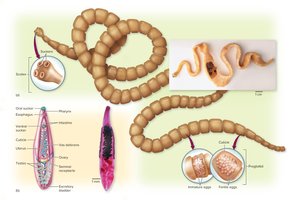
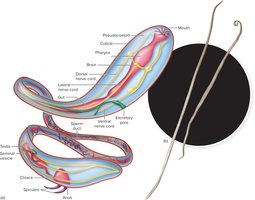
Helminths: Structure, Life Cycle, and Disease
Major Groups of Helminths
Helminths are multicellular parasitic worms, including:
Flatworms (Platyhelminthes): Thin, segmented (cestodes/tapeworms) or unsegmented (trematodes/flukes).
Roundworms (Nematodes): Cylindrical, unsegmented bodies.
Helminth Life Cycle
Helminths have complex life cycles involving eggs, larvae, and adults. Transmission often involves intermediate and definitive hosts. Eggs are released into the environment and may infect new hosts via contaminated food, water, or soil.
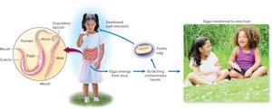
Example: Pinworm Life Cycle
The pinworm (Enterobius vermicularis) is a common intestinal parasite. Its life cycle involves ingestion of eggs, maturation in the intestine, and deposition of eggs around the anus, leading to reinfection and transmission.
Summary Table: Comparison of Eukaryotic Microorganisms
Group | Cell Type | Key Features | Reproduction | Examples |
|---|---|---|---|---|
Fungi | Eukaryotic | Cell wall (chitin), heterotrophic, spores | Asexual/sexual spores | Yeasts, molds |
Protozoa | Eukaryotic | No cell wall, motile, trophozoite/cyst stages | Asexual/sexual | Amoebas, ciliates, flagellates, apicomplexans |
Helminths | Eukaryotic (multicellular) | Complex life cycles, organ systems | Sexual (hermaphroditic or separate sexes) | Tapeworms, flukes, roundworms |
