 Back
Backlec 10:Eukaryotic Microbes and Invertebrate Infectious Agents: Structure, Function, and Pathogenesis
Study Guide - Smart Notes
 Tailored notes based on your materials, expanded with key definitions, examples, and context.
Eukaryotic Microbes
Core Characteristics of Eukaryotic Microbes
Eukaryotic microbes are unicellular or multicellular organisms belonging to the domain Eukarya. Their cells possess a nucleus and specialized organelles, distinguishing them from prokaryotes. These organisms are fundamental to ecological systems and human health.
Nucleus: Eukaryotic DNA is enclosed within a nuclear membrane.
Organelles: Specialized structures such as mitochondria (energy production) and chloroplasts (photosynthesis in algae).
Cell Size and Complexity: Eukaryotic cells are generally larger and more complex than prokaryotic cells.
Cell Division: Division occurs via mitosis, ensuring accurate chromosome segregation.
Main Groups of Eukaryotic Microbes
Eukaryotic microbes are classified into four primary categories, each with unique biological roles and characteristics.
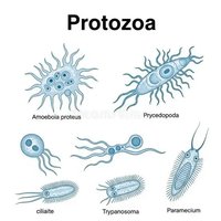
Protozoa: Unicellular, motile organisms, often pathogenic (e.g., Trypanosoma, Giardia).
Algae: Photosynthetic organisms, crucial for oxygen production and aquatic food webs.
Microscopic Fungi: Includes yeasts and molds, important in decomposition and biotechnology.
Other Eukaryotes: Includes less common groups such as slime molds.

Importance of Eukaryotic Microbes
Eukaryotic microbes play essential roles in ecosystems, industry, and medicine.
Ecological Balance: Algae produce oxygen and form the base of aquatic food chains.
Decomposition: Fungi recycle nutrients by breaking down organic matter.
Human Health: Some are beneficial (yeast in bread), others cause diseases (malaria, giardiasis).
Biotechnology: Used in production of medicines, biofuels, and fermented foods.
Invertebrate Infectious Agents
Definition and Types
Invertebrate infectious agents are animals without a backbone that either cause disease directly or act as vectors for pathogens. Their role in disease transmission is significant in both human and veterinary medicine.
Direct Parasitic Agents: Helminths (worms), ectoparasites (lice, fleas, mites), and mollusks (snails).
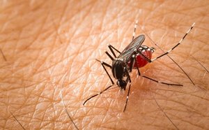
Vectors of Infection: Arthropods (mosquitoes, ticks, flies) transmit pathogens; mechanical vectors (houseflies, cockroaches) carry pathogens without internal development.
Pathogens of Invertebrates: Microbes infecting invertebrates, used in biological pest control.

Flagellated Protozoa: Genus Giardia
Overview and Life Cycle
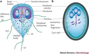
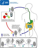
Giardia is a flagellated protozoan parasite that infects the small intestine of vertebrates, causing giardiasis. It alternates between two forms: trophozoite (active) and cyst (infectious, resistant).
Trophozoite: Pear-shaped, two nuclei, four pairs of flagella, ventral disc for attachment.
Cyst: Oval, thick wall, four nuclei, resistant to environmental stress.

Giardia duodenalis: Host Range and Transmission
Giardia duodenalis infects a wide range of hosts, including humans and animals. Transmission occurs via ingestion of cysts from contaminated water, food, or surfaces.
Excystation: Cysts release trophozoites in the small intestine.
Multiplication: Trophozoites multiply by binary fission.
Encystation: Trophozoites form cysts in the colon, which are excreted in stool.


Pathology of Giardiasis
Giardiasis manifests as severe diarrhea, malabsorption, weight loss, and mild intestinal injury. Trophozoites disrupt epithelial cell junctions and brush border enzymes, affecting gastrointestinal motility.
Symptoms: Diarrhea, malnutrition, weight loss.
Mechanism: Adherence to intestinal mucosa via ventral disc.
Diagnosis of Giardiasis
Diagnosis relies on stool antigen detection assays, nucleic acid amplification tests (NAAT), and microscopy. Multiple stool samples increase sensitivity due to intermittent shedding.
Antigen/NAAT: High sensitivity.
Microscopy: Essential for confirmation, especially with multiple samples.

Treatment of Giardiasis
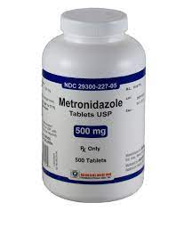
Initial management includes oral rehydration and, if necessary, intravenous fluids. Metronidazole is the first-line drug, with alternatives for special populations.
Metronidazole: First-line therapy; caution in pregnancy.
Other drugs: Tinidazole, nitazoxanide, mebendazole, albendazole, paromomycin.
Epidemiology of Giardiasis
Giardia is prevalent worldwide, with over 1 million cases annually in the United States. Risk factors include childcare settings, travel to areas with poor sanitation, recreational water exposure, and contact with infected animals.
High-risk groups: Children, travelers, swimmers, animal handlers.

Vectorborne Viral Diseases: Zika Virus
Genus Orthoflavivirus: Zika Virus
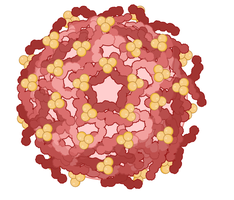
Zika virus is a mosquito-borne, single-stranded positive-sense RNA virus in the Flaviviridae family. Other flaviviruses include dengue, yellow fever, and West Nile virus.
Structure: Enveloped, icosahedral virion.
Transmission: Primarily by Aedes aegypti and Aedes albopictus mosquitoes.
Zika Virus Pathogenesis
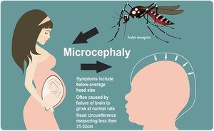
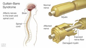
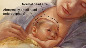
Zika virus targets neural progenitor cells in the fetal brain, causing microcephaly and other neurological defects. In adults, it is associated with Guillain-Barré Syndrome (GBS), a disorder of nerve demyelination.
Microcephaly: Abnormally small head/brain in infants.
GBS: Immune-mediated nerve damage, muscle weakness, paralysis.
Zika Virus Transmission
Zika virus is transmitted through mosquito bites, mother-to-fetus, sexual contact, and rarely, blood transfusion. The virus persists in semen longer than other body fluids.
Mosquito bites: Aedes aegypti and Aedes albopictus.
Vertical transmission: Mother to fetus.
Sexual transmission: All forms of sexual contact, including asymptomatic carriers.
Blood transfusion: Rare, but possible.

Zika Virus Transmission Cycles
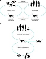
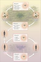
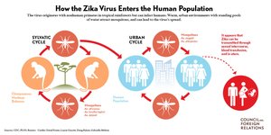
Zika virus transmission occurs in two main cycles: sylvatic (rural/enzootic) and urban. The sylvatic cycle involves non-human primates and forest mosquitoes, while the urban cycle involves human-mosquito-human transmission in populated areas.
Sylvatic cycle: Non-human primates and forest mosquitoes; humans are spillover hosts.
Urban cycle: Human-mosquito-human transmission; mosquitoes breed in domestic containers.

Zika Virus Vectors
The primary vectors for Zika virus are Aedes aegypti and Aedes albopictus mosquitoes. These species are responsible for both sylvatic and urban transmission cycles.
Primary vector: Aedes aegypti
Secondary vector: Aedes albopictus

Zika Virus Pathogenesis and Tissue Tropism
Zika virus exhibits tissue tropism, infecting various organs and fluids, including the brain, placenta, eye, testis, uterus, and body fluids.
Brain: Neural progenitor cells, astrocytes.
Placenta: Trophoblasts, endothelial cells.
Eye: Ganglion cells, optic nerve.
Testis: Leydig and Sertoli cells.
Uterus/Vagina: Epithelial cells.
Body fluids: Blood, semen, saliva, urine.
Summary Table: Eukaryotic Microbes and Invertebrate Infectious Agents
Category | Key Features | Examples | Role in Disease |
|---|---|---|---|
Protozoa | Unicellular, motile, nucleus, organelles | Giardia, Trypanosoma | Giardiasis, sleeping sickness |
Algae | Photosynthetic, aquatic, nucleus, chloroplasts | Green algae, diatoms | Oxygen production, food webs |
Fungi | Decomposers, nucleus, mitochondria | Yeast, molds | Decomposition, fermentation, disease |
Helminths | Multicellular, parasitic | Tapeworms, roundworms | Direct infection (e.g., ascariasis) |
Arthropod Vectors | Insects, ticks, transmit pathogens | Mosquitoes, flies | Vectorborne diseases (malaria, Zika) |
Key Equations and Concepts
Binary Fission (Giardia):
Excystation:
Review Questions
Parasitic flagellated protozoa that possess a kinetoplast are primarily classified under which group? Answer: C. Kinetoplastida
Which of the following diseases is caused by a kinetoplastid protozoan transmitted by an insect vector? Answer: C. African sleeping sickness
Which distinguishing feature is characteristic of kinetoplastid protozoa but not Giardia or Trichomonas? Answer: C. Presence of a kinetoplast containing mitochondrial DNA
Zika virus belongs to which family? Answer: B. Flaviviridae
The main mosquito vector of Zika virus is: Answer: C. Aedes aegypti
In adults, Zika virus infection has been associated with: Answer: B. Guillain-Barré Syndrome
The primary mode of Zika virus transmission is: Answer: B. Bite of infected Aedes mosquitoes
Which of the following can transmit Zika virus sexually? Answer: C. Even asymptomatic individuals
The sylvatic cycle of Zika virus involves transmission between: Answer: B. Non-human primates and forest mosquitoes
