 Back
BackFunctional Anatomy of Prokaryotic and Eukaryotic Cells: Microbiology Study Notes
Study Guide - Smart Notes
 Tailored notes based on your materials, expanded with key definitions, examples, and context.
Functional Anatomy of Prokaryotic and Eukaryotic Cells
Introduction
This chapter explores the structural and functional differences between prokaryotic and eukaryotic cells, focusing on their components, cell walls, membranes, and specialized structures. Understanding these differences is fundamental to microbiology, as it underpins the classification, physiology, and pathogenicity of microorganisms.
Components of All Cells
Universal Cell Structures
Plasma (Cell) Membrane: Separates the living cell from its environment, controls entry and exit of substances.
Chromosomes: DNA molecules carrying hereditary information.
Ribosomes: Sites of protein synthesis.
Cytosol: Semi-fluid substance inside the cell membrane.
Prokaryotic vs. Eukaryotic Cells
Key Differences
Prokaryotes: No nucleus, one circular chromosome, no histones, no membrane-bound organelles, cell wall (peptidoglycan in bacteria, pseudomurein in Archaea), divide by binary fission.
Eukaryotes: True nucleus, paired linear chromosomes, histones, membrane-bound organelles, cell wall (chitin in fungi, cellulose in plants), divide by mitosis.
Prokaryotic Cell Structure
Shapes and Arrangements
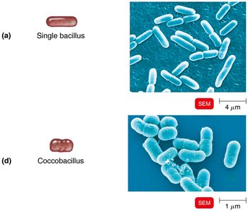
Bacteria exhibit various shapes and arrangements, which are important for identification and classification.
Coccus: Spherical
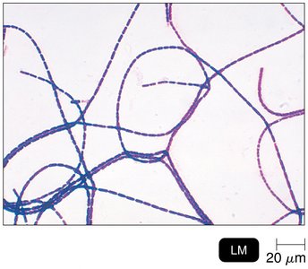
Bacillus: Rod-shaped
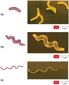
Spiral: Includes vibrio, spirillum, and spirochete forms
Arrangements: Diplococci (pairs), streptococci (chains), staphylococci (clusters), diplobacilli (pairs), streptobacilli (chains)
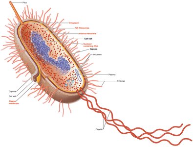
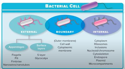
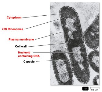
Generalized Prokaryotic Cell Structure
Prokaryotic cells have several key structures that contribute to their function and survival.
Capsule
Cell wall
Plasma membrane
Cytoplasm
Nucleoid (DNA)
Ribosomes
Plasmid
Fimbriae
Flagella
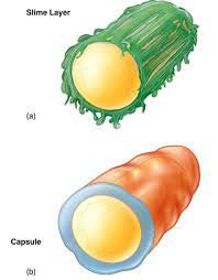
Glycocalyx: Slime Layer and Capsule
Structure and Function
Slime Layer: Loosely organized, promotes adherence, protects from drying, traps nutrients, important in biofilm formation.
Capsule: Highly organized, prevents phagocytosis, thick layer, increases pathogenicity.
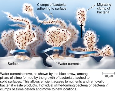
Biofilms
Biofilms are microbial communities that form on surfaces, providing protection and enhanced survival for bacteria.
Form slime or hydrogels
Quorum sensing enables cell-to-cell communication
Advantages: nutrient sharing, resistance to antibiotics and immune system
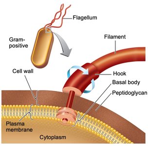
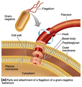
Flagella and Motility
Structure of Prokaryotic Flagellum
Filament: Composed of flagellin, forms a helix
Hook: Connects filament to basal body
Basal Body: Anchors flagellum to cell wall and membrane; structure differs in Gram-positive and Gram-negative bacteria
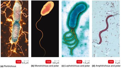
Flagellar Arrangements and Motility
Peritrichous: Flagella distributed over entire cell
Monotrichous: Single flagellum at one pole
Lophotrichous: Tuft of flagella at one pole
Amphitrichous: Flagella at both poles
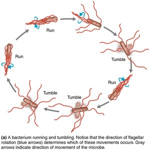
Motility: Bacteria move by "running" and "tumbling"; direction determined by flagellar rotation
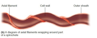
Axial Filaments (Endoflagella)
Found in spirochetes
Anchored at one end, rotation causes corkscrew movement

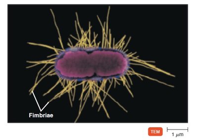
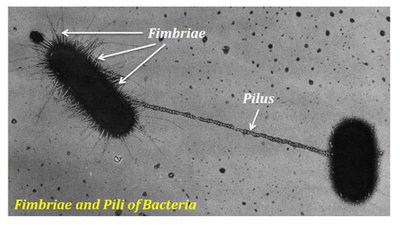
Fimbriae and Pili
Fimbriae
Hairlike appendages for adhesion to surfaces and tissues
Important for colonization and infection
Pili
Rigid tubular structures made of pilin
Assist in attachment, motility (gliding/twitching), and genetic material transfer (conjugation)
Bacterial Cell Wall
Structure and Function
Located outside the plasma membrane
Prevents osmotic lysis, protects cell, contributes to pathogenicity
Composed of peptidoglycan (in bacteria)
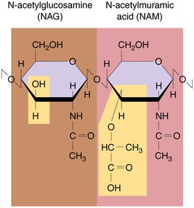
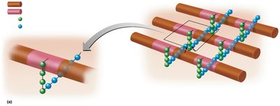
Peptidoglycan Structure
Polymer of disaccharides: N-acetylglucosamine (NAG) and N-acetylmuramic acid (NAM)
Rows of carbohydrates linked by polypeptides
Provides strength and rigidity
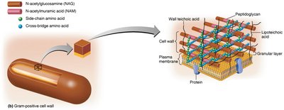
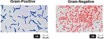
Gram-Positive Cell Wall
Thick peptidoglycan layer
Teichoic acids (wall and lipoteichoic acids) provide antigenic specificity and regulate cation movement
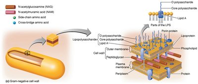
Gram-Negative Cell Wall
Thin peptidoglycan layer
Outer membrane contains lipopolysaccharides (LPS), lipoproteins, and phospholipids
Periplasmic space between outer and plasma membranes
No teichoic acids
LPS contains O polysaccharide (antigen), core polysaccharide (stability), and Lipid A (endotoxin)
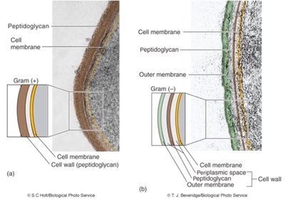
Gram-Positive vs. Gram-Negative Cell Walls
Feature | Gram-Positive | Gram-Negative |
|---|---|---|
Peptidoglycan | Thick | Thin |
Teichoic acids | Present | Absent |
Outer membrane | Absent | Present |
Periplasmic space | Absent | Present |
Atypical Cell Walls
Special Cases
Mycobacterium tuberculosis: Acid-fast cell wall, waxy lipid (mycolic acid), resistant to chemicals and dehydration
Mycoplasma pneumoniae: Lacks cell wall, pleomorphic, sterols in membrane for protection
Damage to the Cell Wall
Mechanisms
Lysozyme: Enzyme that hydrolyzes peptidoglycan bonds
Penicillin: Inhibits peptide bridge formation in peptidoglycan
Plasma Membrane in Bacteria
Fluid Mosaic Model
Phospholipid bilayer with hydrophilic heads and hydrophobic tails
Integral, transmembrane, and peripheral proteins
Functions: energy reactions (ATP), nutrient processing, transport
Self-sealing, proteins and lipids move freely
Membrane Transport
Passive Transport: No energy required; includes simple diffusion and facilitated diffusion
Active Transport: Requires energy (ATP); moves substances against concentration gradient
Group Translocation: Substance is chemically modified during transport
Osmosis and Solutions
Isotonic: Equal solute concentration inside and outside cell
Hypotonic: Lower solute outside; water enters cell, may cause lysis
Hypertonic: Higher solute outside; water leaves cell, causes plasmolysis
Internal Structures of Prokaryotes
Cytoplasm
80% water, contains proteins, carbohydrates, lipids, ions
Serves as pool for building blocks and energy
Cytoskeleton provides structural support
Nucleoid
Region containing bacterial chromosome (circular, double-stranded DNA)
Plasmids: extrachromosomal DNA, often carry antibiotic resistance or toxin genes
Ribosomes
Sites of protein synthesis
Composed of protein and rRNA
Prokaryotic ribosomes: 70S (50S + 30S subunits)
Inclusions
Inclusion | Function |
|---|---|
Metachromatic granules | Phosphate reserves |
Polysaccharide granules | Energy reserves |
Lipid inclusions | Energy reserves |
Sulfur granules | Energy reserves |
Carboxysomes | CO2 fixation during photosynthesis |
Gas vacuoles | Buoyancy |
Magnetosomes | Iron oxide inclusions |
Endospores
Produced by Gram-positive bacteria (e.g., Bacillus, Clostridium)
Formed under nutrient depletion
Highly resistant to adverse conditions
Germination returns endospore to vegetative state
Made of keratin, can survive millions of years
Functional Anatomy of Eukaryotic Cells
Overview
Eukaryotic cells are highly compartmentalized, with membrane-bound organelles performing specialized functions.
Nucleus: Contains genetic material, site of transcription
Ribosomes: Site of translation; 80S in cytoplasm, 70S in mitochondria/chloroplasts
Endoplasmic Reticulum (ER): Rough ER (protein processing), Smooth ER (lipid synthesis, detoxification)
Golgi Complex: Modifies, sorts, and ships proteins
Lysosomes: Digestion and waste removal
Vacuoles: Storage and structural support (plants)
Mitochondria: ATP production via cellular respiration
Chloroplasts: Photosynthesis in plants and algae
Cytoskeleton: Structural support, movement, organelle positioning
Endosymbiotic Theory
Eukaryotes evolved from symbiotic relationships between larger and smaller prokaryotic cells
Mitochondria and chloroplasts originated from engulfed bacteria
Cytoskeleton Elements
Microfilaments: Actin, cell shape, muscle contraction
Intermediate filaments: Cell shape, organelle stabilization
Microtubules: Tubulin, cell shape, vesicle/chromosome movement, motility
Flagella and Cilia
Both made of microtubules (9+2 arrangement)
Flagella: long, few; Cilia: short, numerous
Provide locomotion or move substances along cell surface
External Structures of Eukaryotic Cells
Glycocalyx
Outermost layer, composed of polysaccharides
Functions: protection, adherence, signal reception
Cell Wall
Found in plants, algae, fungi
Composed of chitin (fungi) or cellulose (plants)
Provides structural support and shape
Cell Membrane
Phospholipid bilayer with embedded proteins
Contains sterols for rigidity
Serves as selectively permeable barrier
Additional info: These notes expand on the original content by providing definitions, examples, and comparisons, ensuring a comprehensive and self-contained study guide for college-level microbiology students.
