 Back
BackFunctional Anatomy of Prokaryotic and Eukaryotic Cells: Study Guide
Study Guide - Smart Notes
 Tailored notes based on your materials, expanded with key definitions, examples, and context.
Cell Structure of Prokaryotes and Eukaryotes
Comparison of Prokaryotic and Eukaryotic Cells
Prokaryotic and eukaryotic cells differ fundamentally in their structural organization, genetic material, and cellular processes. Understanding these differences is essential for microbiology students.
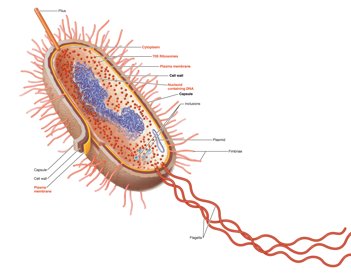
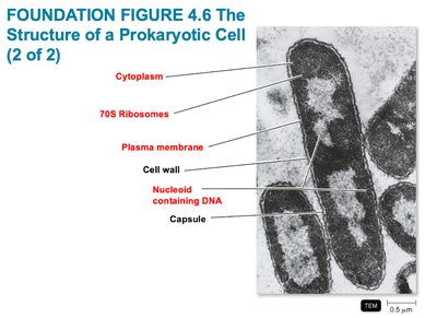
Prokaryotic Cells: Lack a nucleus; DNA is not enclosed by a membrane, usually present as a single circular chromosome. No membrane-bound organelles (e.g., mitochondria, ER, Golgi). Cell wall typically contains peptidoglycan. Divide by binary fission.
Eukaryotic Cells: Possess a true nucleus with DNA enclosed in a nuclear membrane. Multiple linear chromosomes. Membrane-bound organelles are present. Cell walls, when present, are composed of polysaccharides. Divide by mitosis.
Main distinguishing feature: Prokaryotes lack a nucleus, while eukaryotes have a nucleus.
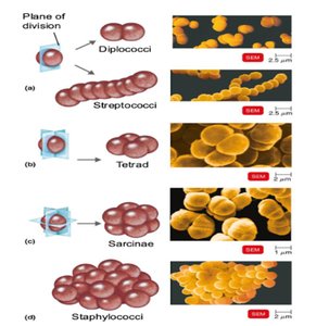
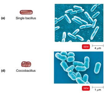
Shapes and Arrangements of Bacteria
Basic Bacterial Shapes and Arrangements
Bacteria exhibit three primary shapes: coccus (spherical), bacillus (rod-shaped), and spiral. Their arrangement is determined by their division patterns and whether cells remain attached after division.
Coccus: Spherical-shaped. Arrangements include diplococci (pairs), streptococci (chains), tetrads (groups of four), sarcinae (cubelike groups of eight), and staphylococci (clusters).
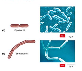
Bacillus: Rod-shaped. Arrangements include single bacillus, diplobacilli (pairs), streptobacilli (chains), and coccobacillus (short, oval rods).
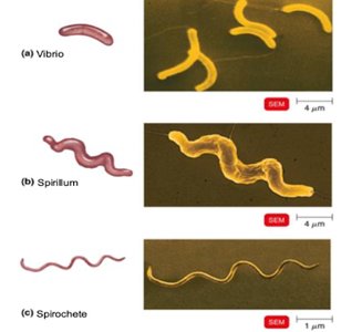
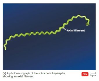
Spiral: Includes vibrio (curved rod), spirillum (rigid spiral), and spirochete (flexible spiral).
Streptococci: Cocci arranged in chains, dividing in one plane and remaining attached.
Glycocalyx: Structure and Function
Bacterial Capsules and Slime Layers
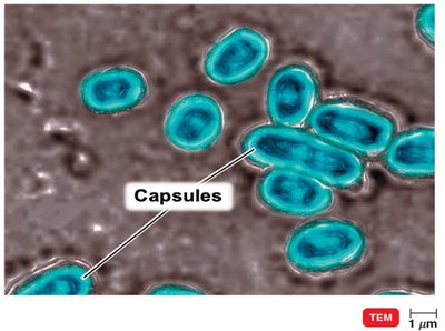
The glycocalyx is an external, viscous, gelatinous layer composed of polysaccharide and/or polypeptide. It exists in two forms: capsule (organized, firmly attached) and slime layer (unorganized, loosely attached).
Capsule: Neatly organized, firmly attached. Contributes to virulence by preventing phagocytosis.
Slime Layer: Unorganized, loosely attached. Facilitates biofilm formation.
Medical Importance: Capsules enable bacteria to evade the immune system, increasing pathogenicity.
Motility and Surface Structures
Flagella, Axial Filaments, Fimbriae, and Pili
Bacteria possess various appendages for motility and attachment. These structures are critical for movement, colonization, and genetic exchange.
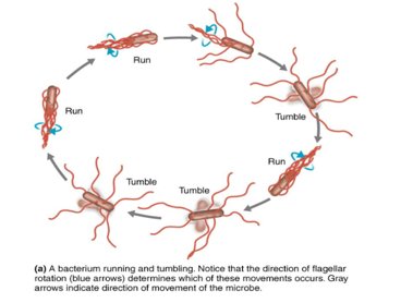
Flagella: Long, filamentous appendages for swimming via rotation. Enable taxis (movement toward/away from stimuli).
Axial Filaments: Also called endoflagella; found in spirochetes. Located between cell wall and outer sheath, causing corkscrew motion.
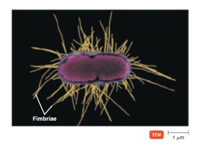
Fimbriae: Short, hairlike structures for attachment to surfaces or host cells. Important for colonization.
Pili: Longer than fimbriae; involved in twitching/gliding motility and conjugation (DNA transfer).
Bacterial Cell Walls
Gram-Positive, Gram-Negative, and Acid-Fast Bacteria
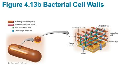
Bacterial cell walls provide structural support and protection. Their composition determines staining properties and antibiotic susceptibility.
Gram-Positive: Thick peptidoglycan, teichoic acids, two rings in flagellar basal body, high susceptibility to penicillin, disrupted by lysozyme.
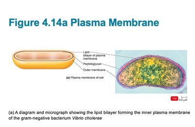
Gram-Negative: Thin peptidoglycan, outer membrane with LPS, four rings in flagellar basal body, low susceptibility to penicillin, resistant to lysozyme.
Acid-Fast: Peptidoglycan with waxy mycolic acid, similar to gram-positive, resistant to staining.
Gram-negative bacteria are harder to treat: Their outer membrane blocks antibiotics, porins limit drug entry, and LPS provides extra protection.
Type | Wall Structure | Antibiotic Susceptibility |
|---|---|---|
Gram-Positive | Thick peptidoglycan, teichoic acid | High |
Gram-Negative | Thin peptidoglycan, LPS, outer membrane | Low |
Acid-Fast | Peptidoglycan + mycolic acid (waxy) | Variable |
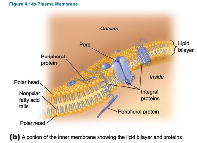
Prokaryotic Plasma Membrane
Structure, Chemistry, and Functions
The prokaryotic plasma membrane is a phospholipid bilayer with embedded proteins, following the fluid mosaic model. It is essential for selective permeability, transport, enzymatic activity, and energy production.
Structure: Phospholipid bilayer, peripheral and integral proteins, self-sealing.
Chemistry: Composed of phospholipids and proteins; proteins act as channels, carriers, and enzymes.
Functions: Selective permeability, ATP production, photosynthesis (chromatophores in some bacteria).
Agents causing injury: Alcohols, quaternary ammonium, polymyxin antibiotics, polypeptide antibiotics.
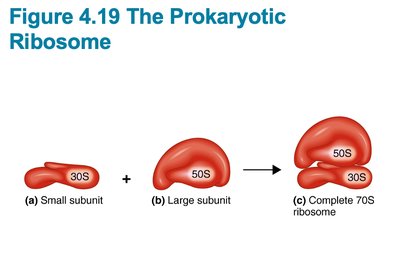
Nucleoid and Ribosomes
Functions and Location in Prokaryotes
The nucleoid contains the bacterial chromosome and stores genetic information, controlling cell structure, metabolism, and reproduction. Ribosomes are the site of protein synthesis, translating mRNA into proteins.
Nucleoid: Single, circular, double-stranded DNA; not surrounded by a nuclear membrane; attached to plasma membrane.
Ribosomes: 70S ribosomes (50S + 30S subunits); site of protein synthesis; essential for enzyme production and metabolism.
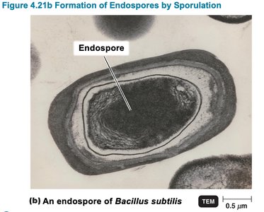
Endospores, Sporulation, and Germination
Survival Structures in Bacteria
Endospores are highly resistant survival structures formed by certain bacteria (e.g., Bacillus, Clostridium) under harsh conditions. Sporulation is the process of endospore formation, while germination is the return to a vegetative cell when conditions improve.
Endospores: Resist desiccation, heat, chemicals, radiation; not reproductive structures.
Sporulation: Occurs when nutrients are depleted; cell forms a thick protective structure around DNA.
Germination: Endospore absorbs water and resumes normal metabolism when conditions become favorable.
Ribosome Structure and Antibiotic Action
Comparison of Prokaryotic and Eukaryotic Ribosomes
Prokaryotic ribosomes are 70S (30S + 50S), while eukaryotic ribosomes are 80S (40S + 60S). Antibiotics like erythromycin target the 50S subunit of prokaryotic ribosomes, inhibiting protein synthesis and bacterial growth, but have minimal effect on eukaryotic ribosomes.
Prokaryotic Ribosomes: 70S, found in cytoplasm.
Eukaryotic Ribosomes: 80S, found in cytoplasm and rough ER.
Erythromycin: Binds 50S subunit, inhibits protein synthesis in bacteria; generally safe for human cells, but may affect mitochondria (which contain 70S ribosomes).
Endosymbiotic Theory of Eukaryotic Evolution
Evidence for Prokaryotic Origins of Mitochondria
The endosymbiotic theory proposes that eukaryotic cells evolved from prokaryotic cells through symbiosis. Mitochondria share several features with bacteria, supporting this theory.
Size and Shape: Mitochondria are similar to bacteria.
DNA: Mitochondria contain circular DNA, like bacteria.
Replication: Mitochondria replicate independently by binary fission.
Ribosomes: Mitochondria have 70S ribosomes, similar to prokaryotes.
Protein Synthesis: Mitochondrial protein synthesis resembles bacterial processes; antibiotics affecting bacterial ribosomes also affect mitochondria.
