Back
BackFunctional Anatomy of Prokaryotic and Eukaryotic Cells: Microbiology Study Notes
Study Guide - Smart Notes
 Tailored notes based on your materials, expanded with key definitions, examples, and context.
Functional Anatomy of Prokaryotic and Eukaryotic Cells
Overview: Prokaryotic vs. Eukaryotic Cells
This section introduces the fundamental differences between prokaryotic and eukaryotic cells, which are the two major cell types found in the microbial world. Understanding these differences is essential for studying microbial structure, function, and classification.
Prokaryotes: Organisms whose cells lack a true nucleus and membrane-bound organelles. Includes Bacteria and Archaea.
Eukaryotes: Organisms whose cells have a true nucleus enclosed by a nuclear membrane and possess membrane-bound organelles. Includes Fungi, Protozoa, Algae, and Helminths.
Key Differences:
Prokaryotes: One circular chromosome (not in a membrane), no histones, no organelles, cell walls (peptidoglycan in bacteria, pseudomurein in archaea), divide by binary fission.
Eukaryotes: Paired chromosomes in a nuclear membrane, histones present, organelles present, polysaccharide cell walls (when present), divide by mitosis.
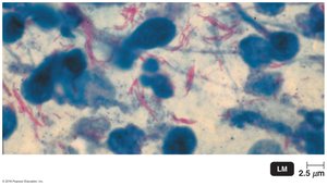
Size, Shape, and Arrangement of Bacterial Cells
General Characteristics
Bacteria exhibit a variety of shapes and arrangements, which are important for identification and classification.
Average Size: 0.2 to 2.0 μm in diameter, 2 to 8 μm in length.
Monomorphic: Most bacteria maintain a single shape.
Pleomorphic: Some bacteria can vary in shape.
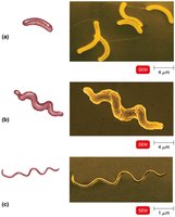
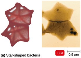
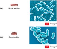
Common Shapes
Bacillus: Rod-shaped
Coccus: Spherical-shaped
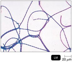
Spiral: Includes vibrio (comma-shaped), spirillum (rigid spiral), and spirochete (flexible spiral)
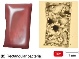
Other Shapes: Star-shaped, rectangular
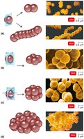
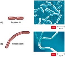
Arrangements
Pairs: Diplococci, diplobacilli
Chains: Streptococci, streptobacilli
Clusters: Staphylococci
Groups of Four: Tetrads
Cubelike Groups of Eight: Sarcinae
Example: Bacillus anthracis
Bacillus anthracis is a rod-shaped, Gram-positive bacterium that causes anthrax.
Structure of a Prokaryotic Cell
Prokaryotic cells have a simple structure but possess specialized features that contribute to their survival and pathogenicity.
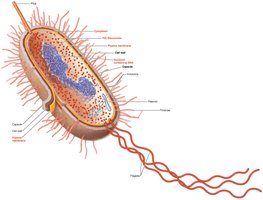
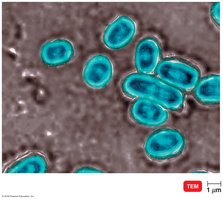
Glycocalyx
Definition: A viscous, gelatinous polymer external to the cell wall, composed of polysaccharide and/or polypeptide.
Types:
Capsule: Neatly organized and firmly attached.
Slime Layer: Unorganized and loosely attached.
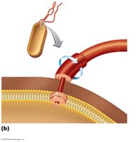
Functions: Contributes to virulence by preventing phagocytosis and aiding in biofilm formation.
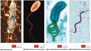
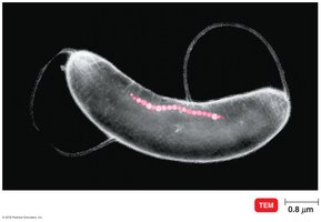
Flagella
Definition: Long, filamentous appendages that propel bacteria.
Structure: Composed of three parts: filament (outermost), hook (connects filament to cell), and basal body (anchors flagellum).
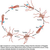
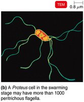
Function: Motility (movement toward/away from stimuli, called taxis); flagella rotate to produce "runs" and "tumbles".
Antigenic Properties: Flagella proteins (H antigens) are used to distinguish bacterial serovars.
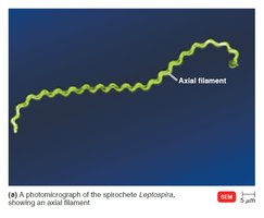
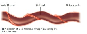
Archaella and Axial Filaments
Archaella: Motility structures in archaea, composed of archaellins (glycoproteins), rotate like flagella.
Axial Filaments (Endoflagella): Found in spirochetes, anchored at one end, rotation causes corkscrew movement.
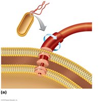
Fimbriae and Pili
Fimbriae: Hairlike appendages for attachment to surfaces and other cells.
Pili: Involved in motility (gliding, twitching) and DNA transfer (conjugation pili).

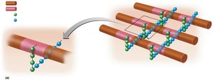
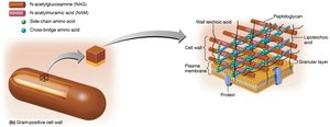
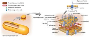
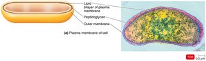
The Cell Wall
The bacterial cell wall is a rigid structure that maintains cell shape, prevents osmotic lysis, and contributes to pathogenicity. Its composition varies between Gram-positive and Gram-negative bacteria.
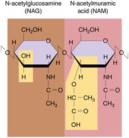
Peptidoglycan Structure
Peptidoglycan: A polymer of repeating disaccharides (N-acetylglucosamine, NAG, and N-acetylmuramic acid, NAM) cross-linked by polypeptides.
Gram-Positive Cell Walls
Thick peptidoglycan layer
Teichoic acids: Lipoteichoic acid links cell wall to plasma membrane; wall teichoic acid links peptidoglycan.
2 rings in basal body of flagella
Produce exotoxins
High susceptibility to penicillin
Disrupted by lysozyme
Gram-Negative Cell Walls
Thin peptidoglycan layer
Outer membrane: Contains lipopolysaccharide (LPS), lipoproteins, and phospholipids.
Periplasmic space: Contains peptidoglycan.
LPS: O polysaccharide (antigenic), Lipid A (endotoxin).
4 rings in basal body of flagella
Produce endotoxins and exotoxins
Low susceptibility to penicillin
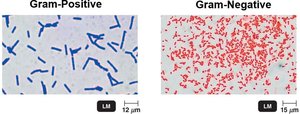
Gram Stain Mechanism
Gram-positive: Alcohol dehydrates peptidoglycan, crystal violet-iodine (CV-I) complexes are retained, cells appear purple.
Gram-negative: Alcohol dissolves outer membrane, CV-I washes out, cells are colorless until counterstained with safranin (appear red/pink).
Atypical Cell Walls
Acid-fast cell walls: Like Gram-positive but with waxy mycolic acid (e.g., Mycobacterium, Nocardia).
Mycoplasmas: Lack cell walls, have sterols in plasma membrane.
Archaea: May lack cell walls or have walls of pseudomurein (lack NAM and D-amino acids).
Damage to the Cell Wall
Lysozyme: Hydrolyzes bonds in peptidoglycan.
Penicillin: Inhibits peptide bridges in peptidoglycan.
Protoplast: Wall-less Gram-positive cell.
Spheroplast: Wall-less Gram-negative cell.
L forms: Wall-less cells that swell into irregular shapes.
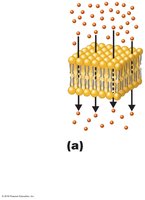
The Plasma (Cytoplasmic) Membrane
The plasma membrane is a selectively permeable barrier composed of a phospholipid bilayer with embedded proteins. It plays a critical role in transport, energy production, and cell signaling.
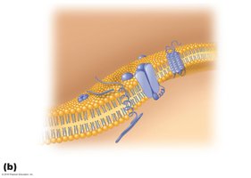
Structure: Fluid Mosaic Model
Membrane is as viscous as olive oil.
Proteins move freely for various functions.
Phospholipids rotate and move laterally.
Self-sealing property.
Functions
Selective permeability: Allows passage of some molecules but not others.
ATP production: Contains enzymes for energy generation.
Photosynthetic pigments: Found on foldings called chromatophores in some bacteria.
Damage to the Membrane
Alcohols, detergents, and antibiotics (e.g., polymyxin) can disrupt the membrane, causing leakage of cell contents.
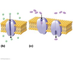
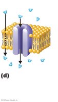
Movement of Materials Across Membranes
Cells transport substances across their membranes using passive and active processes.
Passive Processes
Simple diffusion: Movement of solute from high to low concentration until equilibrium is reached.
Facilitated diffusion: Solute combines with a transporter protein; used for ions and larger molecules.
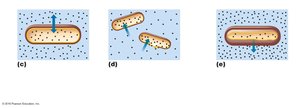
Osmosis: Movement of water across a selectively permeable membrane from high to low water concentration, via lipid layer or aquaporins.
Osmotic pressure: Pressure needed to stop water movement across the membrane.
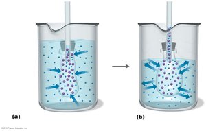
Isotonic solution: Equal solute concentrations inside and outside cell; no net water movement.
Hypotonic solution: Lower solute outside; water enters cell, may cause lysis.
Hypertonic solution: Higher solute outside; water leaves cell, causing plasmolysis.
Active Processes
Active transport: Requires transporter protein and ATP; moves substances against concentration gradient.
Group translocation: Requires transporter protein and phosphoenolpyruvic acid (PEP); substance is chemically altered during transport.
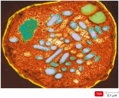
Cytoplasm and Internal Structures
The cytoplasm is the substance inside the plasma membrane, containing water, proteins, carbohydrates, lipids, ions, and the cytoskeleton.
The Nucleoid
Bacterial chromosome: Circular DNA containing genetic information.
Plasmids: Extrachromosomal DNA elements carrying non-essential genes (e.g., antibiotic resistance).
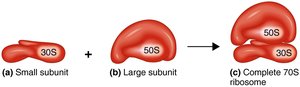
Ribosomes
Function: Sites of protein synthesis.
Structure: Composed of protein and rRNA; 70S (50S + 30S subunits) in prokaryotes.
Inclusions
Metachromatic granules (volutin): Phosphate reserves.
Polysaccharide granules, lipid inclusions, sulfur granules: Energy reserves.
Carboxysomes: Contain RuBisCO for CO2 fixation in photosynthesis.
Gas vacuoles: Protein-covered cylinders for buoyancy.
Magnetosomes: Iron oxide inclusions for orientation in magnetic fields.
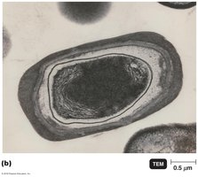
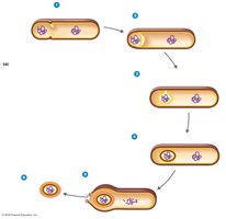
Endospores
Definition: Resting, highly resistant cells formed by certain bacteria (e.g., Bacillus, Clostridium) when nutrients are depleted.
Resistance: Endospores withstand desiccation, heat, chemicals, and radiation.
Sporulation: Process of endospore formation.
Germination: Return of endospore to vegetative state.