 Back
BackFunctional Anatomy of Prokaryotic and Eukaryotic Cells: Structure, Function, and Comparison
Study Guide - Smart Notes
 Tailored notes based on your materials, expanded with key definitions, examples, and context.
Functional Anatomy of Prokaryotic and Eukaryotic Cells
Introduction
This chapter explores the structural and functional differences between prokaryotic and eukaryotic cells, focusing on their cellular components, arrangements, and adaptations. Understanding these differences is fundamental to microbiology, as it underpins microbial classification, physiology, and pathogenicity.
Comparing Prokaryotic and Eukaryotic Cells
Basic Definitions and Overview
Prokaryote: Organisms whose cells lack a true nucleus and membrane-bound organelles. The term derives from Greek for "prenucleus." Includes Bacteria and Archaea.
Eukaryote: Organisms with cells containing a true nucleus enclosed by a nuclear membrane and various membrane-bound organelles. The term means "true nucleus." Includes Fungi, Algae, Protozoa, plants, and animals.
Feature | Prokaryote | Eukaryote |
|---|---|---|
Chromosomes | Usually one circular, not in a membrane | Paired, in nuclear membrane |
Histones | Absent | Present |
Organelles | Absent | Present |
Cell Wall | Peptidoglycan (Bacteria), Pseudomurein (Archaea) | Polysaccharide (when present) |
Division | Binary fission | Mitosis |
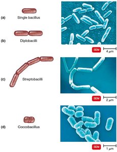
The Size, Shape, and Arrangement of Bacterial Cells
Size and Morphology
Average size: 0.2–2.0 μm in diameter, 2–8 μm in length.
Most bacteria are monomorphic (single shape); some are pleomorphic (variable shapes).
Common Shapes
Bacillus: Rod-shaped
Coccus: Spherical
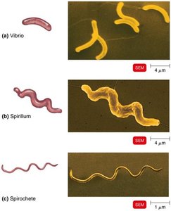
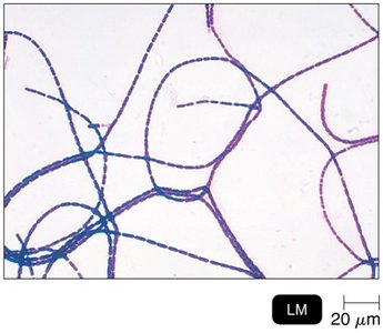
Spiral: Includes Vibrio (comma-shaped), Spirillum (rigid spiral), Spirochete (flexible spiral)
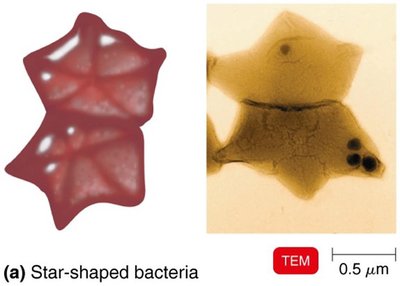
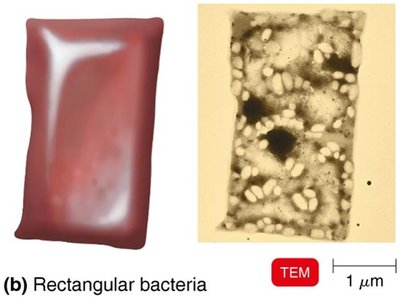
Other: Star-shaped, rectangular
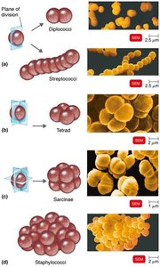
Arrangements
Pairs: Diplococci, diplobacilli
Chains: Streptococci, streptobacilli
Clusters: Staphylococci
Groups of four: Tetrads
Cubelike groups of eight: Sarcinae
Structure of a Prokaryotic Cell
Generalized Structure
Prokaryotic cells contain a cell wall, plasma membrane, cytoplasm, nucleoid, ribosomes, and various external structures such as flagella, fimbriae, and pili.
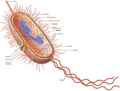
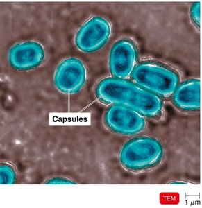
Glycocalyx
Structure and Function
External to the cell wall; viscous and gelatinous.
Composed of polysaccharide and/or polypeptide.
Types: Capsule (organized, firmly attached), Slime layer (unorganized, loose).
Functions: Increases virulence by preventing phagocytosis, aids in adherence to surfaces, and forms biofilms.
Examples: Bacillus anthracis, Streptococcus pneumoniae, Klebsiella pneumoniae (capsule); Streptococcus mutans, Vibrio cholerae (biofilm formation).
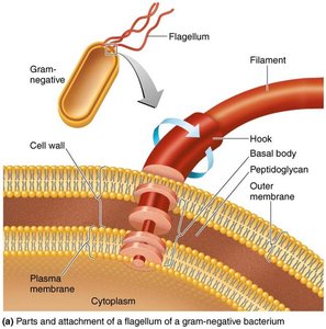
Flagella
Structure and Function
Filamentous appendages for motility, composed of flagellin protein.
Three parts: Filament (outermost), Hook (attaches filament), Basal body (anchors to cell wall and membrane).
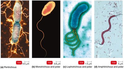
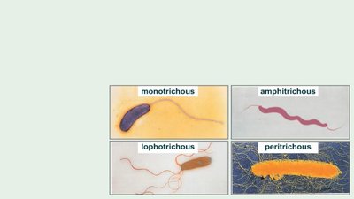
Arrangements
Monotrichous: Single flagellum
Lophotrichous: Tuft at one end
Amphitrichous: One or more at both ends
Peritrichous: Distributed over entire cell
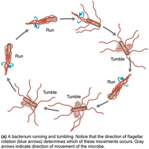
Function in Motility and Identification
Enable movement toward/away from stimuli (taxis).
Flagella proteins (H antigens) are used to distinguish serovars (e.g., E. coli O157:H7).
Other Surface Structures
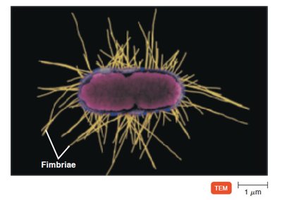
Fimbriae and Pili
Fimbriae: Hairlike appendages for attachment and biofilm formation (e.g., Neisseria gonorrhoeae, E. coli O157).
Pili: Involved in motility (gliding, twitching) and DNA transfer (conjugation pili).
The Cell Wall
Composition and Function
Prevents osmotic lysis, protects membrane, and contributes to pathogenicity.
Composed of peptidoglycan in bacteria: polymer of N-acetylglucosamine (NAG) and N-acetylmuramic acid (NAM) linked by polypeptides.
Target for antibiotics (e.g., penicillin inhibits peptide cross-bridges).
Gram-Positive vs. Gram-Negative Cell Walls
Feature | Gram-Positive | Gram-Negative |
|---|---|---|
Peptidoglycan | Thick, many layers | Thin, single layer |
Teichoic acids | Present | Absent |
Outer membrane | Absent | Present (contains LPS) |
Flagella basal body | 2 rings | 4 rings |
Toxins | Exotoxins | Endotoxins & Exotoxins |
Penicillin susceptibility | High | Low |
Gram Stain Mechanism
Crystal violet-iodine complex forms inside cells.
Alcohol dehydrates peptidoglycan in gram-positive cells (retains dye).
Alcohol dissolves outer membrane in gram-negative cells (dye washes out, safranin counterstain applied).
Atypical Cell Walls
Acid-fast bacteria: Thick peptidoglycan, waxy mycolic acid (e.g., Mycobacterium).
Mycoplasmas: Lack cell walls, contain sterols in membrane.
Archaea: Wall-less or walls of pseudomurein (lack NAM and D-amino acids).
Damage to the Cell Wall
Lysozyme: Hydrolyzes peptidoglycan bonds (especially in gram-positive bacteria).
Penicillin: Inhibits peptide bridge formation in peptidoglycan.
Protoplast: Wall-less gram-positive cell; Spheroplast: Wall-less gram-negative cell; L forms: Irregular, wall-less cells.
The Plasma (Cytoplasmic) Membrane
Structure
Phospholipid bilayer with embedded proteins (integral, peripheral, transmembrane).
Some proteins and lipids have attached carbohydrates (glycoproteins, glycolipids).
Fluid Mosaic Model
Membrane is fluid, allowing lateral movement of proteins and lipids.
Self-sealing and selectively permeable.
Functions
Selective permeability: controls entry/exit of substances.
Contains enzymes for ATP production.
Photosynthetic pigments (chromatophores) in some bacteria.
Destruction by Antimicrobial Agents
Alcohols, detergents, and antibiotics (e.g., polymyxin) can damage the membrane, causing leakage of cell contents.
Movement of Materials Across Membranes
Passive Processes
Simple diffusion: Movement from high to low concentration until equilibrium is reached.
Facilitated diffusion: Transport proteins help move substances down their concentration gradient.
Osmosis: Net movement of water across a selectively permeable membrane.
Osmotic pressure: Pressure needed to stop water movement.
Isotonic: Equal solute concentrations; Hypotonic: Lower outside; Hypertonic: Higher outside.
Active Processes
Active transport: Uses ATP and transporter proteins to move substances against their gradient.
Group translocation: Substance is chemically altered during transport (requires PEP).
Cytoplasm and Internal Structures
Cytoplasm
Thick, aqueous, elastic substance inside the plasma membrane.
Contains DNA (nucleoid), ribosomes, inclusions, and cytoskeleton.
Nucleoid
Region containing the bacterial chromosome (circular, double-stranded DNA).
No nuclear envelope or histones.
Plasmids: Small, extrachromosomal DNA circles carrying nonessential genes (e.g., antibiotic resistance).
Ribosomes
Sites of protein synthesis; composed of protein and rRNA.
Prokaryotic ribosomes: 70S (50S + 30S subunits).
Target for antibiotics (e.g., streptomycin, gentamicin).
Inclusions
Reserve deposits for nutrients (e.g., phosphate, polysaccharides, lipids, sulfur).
Specialized inclusions: Carboxysomes (photosynthesis), gas vacuoles (buoyancy), magnetosomes (iron oxide).
Endospores
Resting, highly resistant cells formed by Bacillus and Clostridium when nutrients are scarce.
Resistant to desiccation, heat, chemicals, and radiation.
Sporulation: Formation of endospores; Germination: Return to vegetative state.
Eukaryotic Cell Structures
Flagella and Cilia
Projections for locomotion or moving substances along the cell surface.
Flagella: Long, few; Cilia: Short, numerous.
Both have a "9+2" arrangement of microtubules (tubulin protein).
Movement is wavelike (unlike rotary movement in prokaryotes).
Cell Wall and Glycocalyx
Cell wall: Present in plants (cellulose), fungi (chitin), algae (various polysaccharides).
Glycocalyx: Carbohydrates bonded to proteins/lipids; found in animal cells, aids in cell recognition and attachment.
Plasma Membrane
Similar phospholipid bilayer structure as prokaryotes.
Contains sterols (complex lipids) and carbohydrates (for attachment and recognition).
Functions: Selective permeability, transport, endocytosis (phagocytosis, pinocytosis, receptor-mediated).
Cytoplasm and Organelles
Cytosol: Fluid portion; Cytoskeleton: Microfilaments, intermediate filaments, microtubules.
Cytoplasmic streaming: Movement of cytoplasm within the cell.
Ribosomes
80S (60S + 40S subunits) in cytoplasm and on rough ER; 70S in mitochondria and chloroplasts.
Nucleus
Double membrane (nuclear envelope) encloses DNA complexed with histones (chromatin).
Chromatin condenses into chromosomes during cell division.
Endoplasmic Reticulum (ER)
Rough ER: Studded with ribosomes, site of protein synthesis.
Smooth ER: Synthesizes membranes, fats, hormones.
Golgi Complex
Modifies, sorts, and packages proteins from the ER for transport.
Lysosomes and Vacuoles
Lysosomes: Contain digestive enzymes.
Vacuoles: Storage, shape, and food intake.
Mitochondria and Chloroplasts
Mitochondria: Site of ATP production; double membrane, inner folds (cristae), own DNA and 70S ribosomes.
Chloroplasts: Site of photosynthesis; thylakoids with chlorophyll, own DNA and 70S ribosomes.
Other Organelles
Peroxisomes: Oxidize fatty acids, destroy hydrogen peroxide.
Centrosomes: Organize mitotic spindle, contain centrioles.
The Evolution of Eukaryotes
Endosymbiotic Theory
Proposes that eukaryotes evolved when larger prokaryotic cells engulfed smaller ones, which became organelles (mitochondria, chloroplasts).
Evidence: Mitochondria and chloroplasts resemble bacteria in size/shape, have circular DNA, reproduce independently, have 70S ribosomes, and double membranes.
