 Back
BackFunctional Anatomy of Prokaryotic and Eukaryotic Cells: Study Notes
Study Guide - Smart Notes
 Tailored notes based on your materials, expanded with key definitions, examples, and context.
Functional Anatomy of Prokaryotic and Eukaryotic Cells
Introduction
This chapter explores the structural and functional differences between prokaryotic and eukaryotic cells, which are fundamental to understanding microbiology. The content covers cell types, their distinguishing features, and the diversity of bacterial shapes and structures.
Two Kinds of Cells
Classification of Cells
All living organisms are composed of either prokaryotic or eukaryotic cells. These two cell types differ in their structural organization and complexity.
Prokaryotes: Organisms whose cells lack a true nucleus and membrane-bound organelles. Includes Bacteria and Archaea.
Eukaryotes: Organisms whose cells have a true nucleus enclosed by a nuclear membrane and possess membrane-bound organelles. Includes Animals, Plants, Fungi, Algae, and Protozoa.

Cell Structure Overview
Prokaryotic and eukaryotic cells can be visually distinguished by their internal complexity and the presence or absence of a nucleus.

Prokaryote comes from the Greek for "prenucleus." Their DNA is not enclosed within a membrane.
Eukaryote comes from the Greek for "true nucleus." Their DNA is enclosed within a nuclear membrane.
Differences Between Prokaryotic and Eukaryotic Cells
Genetic Material and Internal Structure
Prokaryotes:
DNA is not enclosed in a nuclear membrane; usually a single circular chromosome.
DNA is not associated with histone proteins.
No membrane-enclosed organelles.
Cell wall, if present, is complex.
Eukaryotes:
DNA is enclosed in a nuclear membrane; multiple linear chromosomes.
DNA is associated with histones and non-histone proteins.
Contains membrane-bound organelles (e.g., Golgi complex, mitochondria, lysosomes).
Cell wall, if present, is simple.
Cell Division and Other Features
Prokaryotes:
Divide by binary fission.
Size: 0.2–20 μm in diameter.
Plasma membrane lacks carbohydrates and generally sterols.
No cytoskeleton or cytoplasmic streaming.
Ribosomes are smaller (70S).
Asexual reproduction (may transfer DNA fragments).
Eukaryotes:
Divide by mitosis; sexual reproduction involves meiosis.
Size: 10–100 μm in diameter.
Plasma membrane contains carbohydrates and sterols (serve as receptors).
Cytoskeleton present.
Ribosomes are larger (80S).
The Prokaryotes
General Characteristics
Prokaryotes include Bacteria and Archaea. Some bacteria, such as cyanobacteria, are photosynthetic. Bacterial species are differentiated by:
Morphology (shape)
Chemical composition (detected by staining)
Nutritional requirements
Biochemical activities
Source of energy (sunlight or chemicals)
Shapes of Bacterial Cells
Basic Shapes
Bacteria exhibit a variety of shapes, which are important for identification and classification.
Bacillus: Rod-shaped
Coccus: Spherical
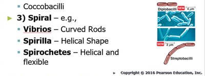
Spiral: Includes Spirillum (rigid, spiral), Vibrio (curved rods), and Spirochete (flexible, helical)
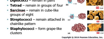
Arrangements of Cocci
Single
Diplococci: Remain in pairs
Tetrad: Groups of four
Sarcinae: Cube-like groups of eight
Streptococci: Chain-like pattern
Staphylococci: Grape-like clusters

Arrangements of Bacilli and Spiral Forms
Bacillus: Single, diplobacilli (pairs), streptobacilli (chains), coccobacilli (short rods)
Spiral: Vibrios (curved rods), spirilla (helical, rigid), spirochetes (helical, flexible)
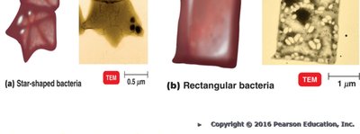
Unusual Shapes
Star-shaped Stella
Square Haloarcula
Monomorphic and Pleomorphic Bacteria
Monomorphic: Most bacteria maintain a single shape, determined by heredity.
Pleomorphic: Some bacteria can have more than one genetically controlled shape (e.g., Rhizobium, Corynebacterium).
Prokaryotic Cell Structure
Diagram of a Prokaryotic Cell
The prokaryotic cell contains several key structures, both internal and external to the cell wall.
Structures External to the Cell Wall
Glycocalyx
A sugar coat on the surface of many cells.
Bacterial glycocalyx is viscous (sticky) and is a gelatinous polymer of polysaccharide and/or polypeptide.
Made inside the cell and secreted to the surface.
If organized and firmly attached, called a capsule.
If unorganized and loose, called a slime layer.
Capsule
Important virulence factor (e.g., Streptococcus pneumoniae is virulent if it has a capsule).
Provides protection from phagocytosis (e.g., Bacillus anthracis).
Allows pathogenic bacteria to attach to surfaces (e.g., Klebsiella in the respiratory tract).
Can serve as a source of nutrition when energy stores are low.
Protects against dehydration.
Capsules are antigenic (contain antigens).
Flagella
Long, filamentous appendages that propel bacteria (motility).
Flagellar protein (H-antigen) is used to distinguish among serovars.
Four arrangements:
Monotrichous: Single polar flagellum
Amphitrichous: Tuft of flagella at both ends
Lophotrichous: Two or more flagella at one pole
Peritrichous: Flagella distributed over the entire cell
Structure of a Prokaryotic Flagellum
Three basic parts:
Filament: Outermost region, constant diameter, composed of flagellin protein arranged in intertwining chains forming a helix around a hollow core.
Hook: Attaches the filament to the cell, wider than the filament, composed of a different protein.
Basal body: Anchors the flagellum to the cell wall and plasma membrane (not shown in the provided images, but important for context).

Additional info: The basal body is a complex structure that differs between Gram-positive and Gram-negative bacteria, anchoring the flagellum and enabling its rotation for motility.
