 Back
BackFunctional Anatomy of Prokaryotic and Eukaryotic Cells: Structure and Function
Study Guide - Smart Notes
 Tailored notes based on your materials, expanded with key definitions, examples, and context.
Functional Anatomy of Prokaryotic and Eukaryotic Cells
Overview: Prokaryotic vs. Eukaryotic Cells
Cells are the fundamental units of life, and in microbiology, they are classified as either prokaryotic or eukaryotic based on structural and functional differences. Understanding these differences is essential for studying microbial physiology, genetics, and taxonomy.
Prokaryotes: Organisms whose cells lack a true nucleus and membrane-bound organelles. Includes Bacteria and Archaea.
Eukaryotes: Organisms with cells containing a true nucleus and various organelles. Includes fungi, algae, protozoa, and helminths.
Feature | Prokaryote | Eukaryote |
|---|---|---|
Chromosomes | One circular, not in membrane | Paired, in nuclear membrane |
Histones | Absent | Present |
Organelles | Absent | Present |
Cell Wall | Peptidoglycan (Bacteria), Pseudomurein (Archaea) | Polysaccharide (when present) |
Division | Binary fission | Mitosis |
The Size, Shape, and Arrangement of Bacterial Cells
Size and Morphology
Bacteria exhibit a variety of shapes and arrangements, which are important for identification and classification.
Average size: 0.2–2.0 μm in diameter, 2–8 μm in length
Monomorphic: Most bacteria have a single, consistent shape
Pleomorphic: Some bacteria can vary in shape
Common Shapes
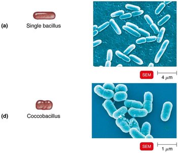
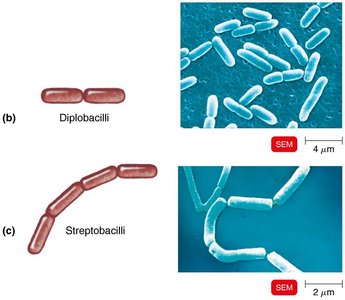
Bacillus: Rod-shaped
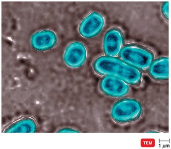
Coccus: Spherical
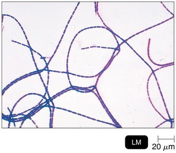
Spiral: Includes vibrio (comma-shaped), spirillum (rigid spiral), and spirochete (flexible spiral)
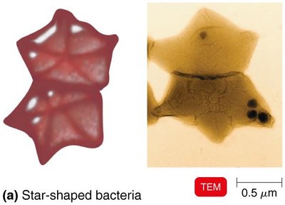
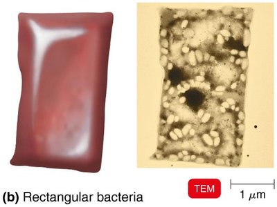
Star-shaped and Rectangular: Rare morphologies

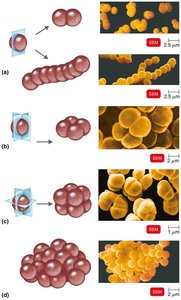
Arrangements
Pairs: Diplococci, diplobacilli
Chains: Streptococci, streptobacilli
Clusters: Staphylococci
Groups of four: Tetrads
Cubelike groups of eight: Sarcinae
Structure of a Prokaryotic Cell
Major Components
Prokaryotic cells have a simple structure but contain all the necessary components for life.
Cell wall: Provides shape and protection
Plasma membrane: Regulates transport
Cytoplasm: Contains enzymes, nutrients, and genetic material
Nucleoid: Region containing the bacterial chromosome
Ribosomes: Sites of protein synthesis
Inclusions: Storage granules
External structures: Glycocalyx, flagella, fimbriae, pili
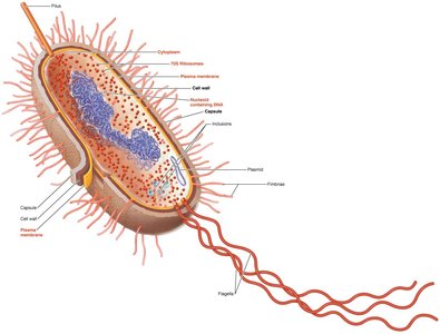
Glycocalyx
Structure and Function
The glycocalyx is a viscous, gelatinous layer external to the cell wall, composed of polysaccharide and/or polypeptide. It exists as either a capsule (organized, firmly attached) or a slime layer (unorganized, loosely attached).
Capsules prevent phagocytosis, contributing to virulence
Extracellular polymeric substance (EPS) helps form biofilms
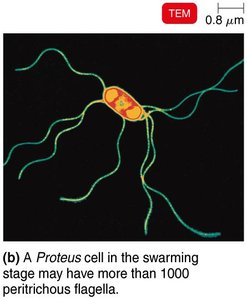
Flagella, Archaella, and Axial Filaments
Flagella
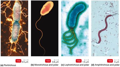
Flagella are long, filamentous appendages that provide motility to bacteria. They are composed of the protein flagellin and consist of three parts: filament, hook, and basal body.
Arrangement: Peritrichous (all over), monotrichous (single, polar), lophotrichous (tuft at one end), amphitrichous (both ends)
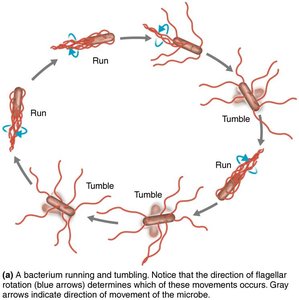
Function: Movement toward/away from stimuli (taxis), "run and tumble" motility
H antigens: Flagellar proteins used for serotyping
Archaella
Archaella are motility structures found in Archaea, composed of glycoproteins called archaellins. They rotate like bacterial flagella but are structurally distinct.
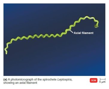
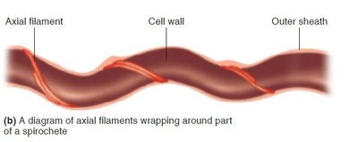
Axial Filaments
Axial filaments, or endoflagella, are found in spirochetes. They are anchored at one end and cause the cell to move in a corkscrew motion.
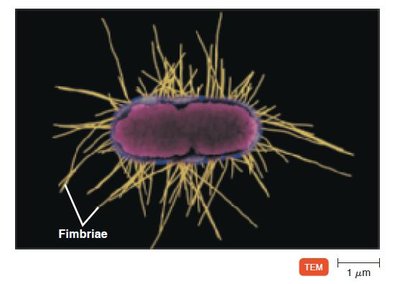
Fimbriae and Pili
Fimbriae
Fimbriae are hairlike appendages that allow bacteria to adhere to surfaces and each other, playing a key role in colonization and biofilm formation.
Pili
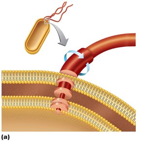
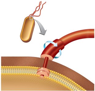
Pili are longer than fimbriae and are involved in motility (gliding, twitching) and the transfer of DNA between cells (conjugation pili).
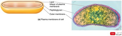
The Cell Wall
Structure and Function
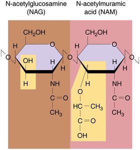
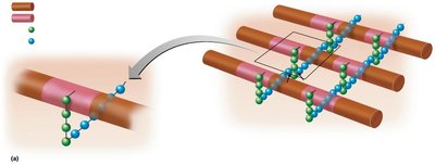
The bacterial cell wall is a rigid structure that prevents osmotic lysis, maintains shape, and contributes to pathogenicity. It is primarily composed of peptidoglycan in bacteria.
Peptidoglycan: Polymer of N-acetylglucosamine (NAG) and N-acetylmuramic acid (NAM) linked by polypeptides
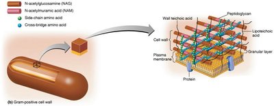
Gram-Positive Cell Walls
Thick peptidoglycan layer
Teichoic acids (lipoteichoic and wall teichoic acids) provide rigidity and antigenic specificity
Two rings in basal body of flagella
Produce exotoxins; highly susceptible to penicillin; disrupted by lysozyme
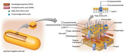
Gram-Negative Cell Walls
Thin peptidoglycan layer
Outer membrane contains lipopolysaccharide (LPS), lipoproteins, and phospholipids
Four rings in basal body of flagella
Produce endotoxins and exotoxins; low susceptibility to penicillin
Porins form channels through the outer membrane
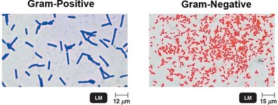
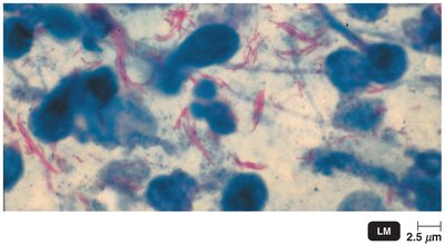
Gram Stain Mechanism
The Gram stain differentiates bacteria based on cell wall structure:
Gram-positive: Alcohol dehydrates peptidoglycan, trapping crystal violet-iodine complex (cells appear purple)
Gram-negative: Alcohol dissolves outer membrane, crystal violet-iodine complex washes out, safranin counterstain colors cells red
Atypical Cell Walls
Acid-fast cell walls: Like Gram-positive but with waxy mycolic acid (e.g., Mycobacterium, Nocardia)
Mycoplasmas: Lack cell walls; have sterols in plasma membrane
Archaea: May lack cell walls or have walls of pseudomurein (lack NAM and D-amino acids)
Damage to the Cell Wall
Lysozyme: Hydrolyzes bonds in peptidoglycan
Penicillin: Inhibits peptide bridges in peptidoglycan
Protoplast: Wall-less Gram-positive cell
Spheroplast: Wall-less Gram-negative cell
L forms: Wall-less cells that swell into irregular shapes
The Plasma (Cytoplasmic) Membrane
Structure
The plasma membrane is a phospholipid bilayer with embedded proteins, following the fluid mosaic model. It is selectively permeable and self-sealing.
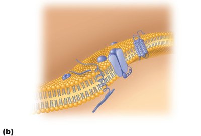
Functions
Selective permeability: Regulates passage of substances
Contains enzymes for ATP production
Photosynthetic pigments may be present on infoldings called chromatophores
Movement of Materials Across Membranes
Passive Processes
Simple diffusion: Movement from high to low concentration until equilibrium is reached
Facilitated diffusion: Solute combines with transporter protein; moves with concentration gradient
Osmosis: Movement of water across a selectively permeable membrane
Osmotic pressure: Pressure needed to stop water movement
Isotonic: Equal solute concentrations
Hypotonic: Lower solute outside; water enters cell (risk of lysis)
Hypertonic: Higher solute outside; water leaves cell (plasmolysis)
Active Processes
Active transport: Requires transporter protein and ATP; moves substances against gradient
Group translocation: Substance is chemically altered during transport (requires PEP)
Cytoplasm and Internal Structures
Cytoplasm
The cytoplasm is the substance inside the plasma membrane, consisting of water, proteins, carbohydrates, lipids, ions, and a cytoskeleton.
Nucleoid
Bacterial chromosome: Circular DNA containing genetic information
Plasmids: Extrachromosomal DNA elements; may carry antibiotic resistance or toxin genes
Ribosomes
Ribosomes are the sites of protein synthesis, composed of protein and rRNA. Prokaryotic ribosomes are 70S (50S + 30S subunits).
Inclusions
Metachromatic granules: Phosphate reserves
Polysaccharide granules: Energy reserves
Lipid inclusions: Energy reserves
Sulfur granules: Energy reserves
Carboxysomes: Contain RuBisCO for CO2 fixation
Gas vacuoles: Maintain buoyancy
Magnetosomes: Iron oxide inclusions; destroy H2O2
Endospores
Formation and Function
Endospores are highly resistant, dormant structures formed by certain bacteria (e.g., Bacillus, Clostridium) when nutrients are depleted. They are resistant to desiccation, heat, chemicals, and radiation.
Sporulation: Process of endospore formation
Germination: Endospore returns to vegetative state
