 Back
BackFunctional Anatomy of Prokaryotic and Eukaryotic Cells: Study Guide
Study Guide - Smart Notes
 Tailored notes based on your materials, expanded with key definitions, examples, and context.
Functional Anatomy of Prokaryotic and Eukaryotic Cells
Overview: Comparing Prokaryotic and Eukaryotic Cells
This chapter explores the structural and functional differences between prokaryotic and eukaryotic cells, focusing on their cellular components, mechanisms of movement, and clinical relevance. Understanding these differences is fundamental for microbiology students, as it underpins microbial classification, physiology, and the basis for antibiotic action.
Prokaryotic cells lack membrane-bound organelles and a nucleus; their cell walls contain peptidoglycan.
Eukaryotic cells possess a membrane-bound nucleus and organelles; their cell walls (if present) do not contain peptidoglycan.
Both cell types share similar chemical compositions and metabolic pathways.
Size, Shape, and Arrangement of Bacterial Cells
Bacteria exhibit a variety of shapes and arrangements, which are important for identification and classification.
Size: Most bacteria are 0.2–2.0 μm in diameter and 2–8 μm in length.
Shapes: Three basic shapes: coccus (spherical), bacillus (rod-shaped), and spiral (twisted).
Pleomorphic bacteria can assume multiple shapes.
Arrangement: Determined by the plane of division and whether cells remain attached after division (e.g., clusters, chains).
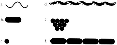
Image description: Diagram showing various bacterial shapes and arrangements, including spirals, rods, cocci, clusters, chains, and spirochetes. This image directly illustrates the diversity of bacterial morphology discussed above.
Structures External to the Cell Wall
Bacteria possess several external structures that contribute to their survival, motility, and pathogenicity.
Glycocalyx: Gelatinous polysaccharide/polypeptide covering (capsule, slime layer, or extracellular polysaccharide).
Capsules: Protect against phagocytosis, aid in adherence, prevent desiccation, and may provide nutrients.
Flagella: Long filamentous appendages for motility; composed of filament, hook, and basal body.
Axial filaments: Endoflagella wrapped around spirochetes, enabling corkscrew movement.
Fimbriae: Short, hair-like structures for adherence to surfaces and other cells.
Pili: Longer than fimbriae; involved in twitching motility and DNA transfer.

Image description: Diagram showing different arrangements of bacterial flagella (monotrichous, lophotrichous, amphitrichous, peritrichous). This image directly supports the explanation of bacterial motility structures.
The Cell Wall: Composition and Characteristics
The bacterial cell wall is a critical structure for protection, shape, and clinical targeting by antibiotics.
Peptidoglycan: Polymer of N-acetylglucosamine (NAG) and N-acetylmuramic acid (NAM) with short amino acid chains.
Gram-positive cell walls: Thick peptidoglycan layer, teichoic acids.
Gram-negative cell walls: Thin peptidoglycan, outer membrane with lipopolysaccharide, lipoprotein, and phospholipid.
Acid-fast cell walls: Mycolic acid layer outside thin peptidoglycan.
Archaea: Pseudomurein cell walls, lack peptidoglycan.
Mycoplasmas: Lack cell walls, resistant to antibiotics targeting cell wall synthesis.
Type | Main Components | Clinical Relevance |
|---|---|---|
Gram-positive | Thick peptidoglycan, teichoic acids | Targeted by penicillin, lysozyme |
Gram-negative | Thin peptidoglycan, outer membrane | Resistant to some antibiotics, contains endotoxin |
Acid-fast | Mycolic acid, thin peptidoglycan | Resistant to many chemicals |
Archaea | Pseudomurein | Not affected by antibiotics targeting peptidoglycan |
Mycoplasma | No cell wall | Resistant to cell wall-targeting antibiotics |
Structures Internal to the Cell Wall
Internal structures are essential for metabolism, genetic information, and survival under adverse conditions.
Plasma membrane: Lipid bilayer with proteins; selectively permeable; site of metabolic reactions.
Cytoplasm: Fluid matrix containing water, molecules, DNA, ribosomes, inclusions, cytoskeleton.
Nucleoid: Region containing bacterial chromosome (DNA); plasmids may be present.
Ribosomes: 70S in prokaryotes; site of protein synthesis; target for antibiotics.
Inclusions: Reserve deposits (e.g., metachromatic granules, glycogen, lipid, sulfur, carboxysomes, gas vacuoles, magnetosomes).
Endospores: Resting structures for survival during adverse conditions; highly resistant.
Movement of Materials Across Membranes
Cells transport materials across membranes using passive and active processes.
Simple diffusion: Movement from high to low concentration until equilibrium.
Facilitated diffusion: Transport via proteins from high to low concentration.
Osmosis: Water movement across membrane toward higher solute concentration.
Active transport: Movement from low to high concentration using energy and transporter proteins.
Group translocation: Chemical modification and transport across membrane using energy.
Key equation for osmosis:
Where is the water potential, determining the direction of water movement.
Eukaryotic Cell Structures
Eukaryotic cells contain specialized organelles and structures for complex functions.
Flagella and cilia: Motility structures; 9+2 microtubule arrangement.
Cell wall: Composition varies (cellulose in algae, chitin in fungi, glucan/mannan in yeast).
Glycocalyx: Strengthens cell, aids attachment.
Plasma membrane: Phospholipid bilayer with proteins, carbohydrates, sterols.
Cytoplasm: Contains cytoskeleton, exhibits cytoplasmic streaming.
Ribosomes: 80S in cytoplasm or on rough ER.
Organelles: Nucleus, ER, Golgi complex, lysosomes, vacuoles, mitochondria, chloroplasts, peroxisomes, centrosomes.
Endosymbiotic Theory of Eukaryotic Evolution
The endosymbiotic theory proposes that eukaryotic cells evolved from symbiotic prokaryotes living inside other prokaryotic cells. Evidence includes similarities between mitochondria/chloroplasts and prokaryotes (e.g., 70S ribosomes, circular DNA, binary fission).
Clinical Applications and Microbiome Exploration
Understanding cell structure informs antibiotic action and the role of eukaryotic microbiota in health. For example, antibiotics targeting 70S ribosomes affect prokaryotes but not human cells. Eukaryotic microbiota may influence immune system maturation.
Summary Table: Prokaryotic vs. Eukaryotic Cell Features
Feature | Prokaryotic Cell | Eukaryotic Cell |
|---|---|---|
Nucleus | No | Yes |
Organelles | No | Yes |
Cell Wall | Peptidoglycan (bacteria) | Cellulose, chitin, glucan, mannan (varies) |
Ribosomes | 70S | 80S |
Flagella | Flagellin protein | Microtubules (9+2 arrangement) |
Case Study: Gram Stain Protocol and Cell Wall Structure
The Gram stain differentiates bacteria based on cell wall structure. Gram-positive bacteria retain crystal violet due to thick peptidoglycan; gram-negative bacteria lose the stain after decolorization and are counterstained with safranin. Errors in protocol can lead to incorrect results.
Correct order: Crystal violet → iodine → decolorizer → safranin.
Common mistakes: Reversing stains, omitting mordant, over-decolorizing.
Additional info: Expanded explanations of cell wall types, transport mechanisms, and clinical relevance were added for completeness and academic context.
