 Back
Back4 Functional Anatomy of Prokaryotic and Eukaryotic Cells: Structure, Function, and Comparison
Study Guide - Smart Notes
 Tailored notes based on your materials, expanded with key definitions, examples, and context.
Comparing Prokaryotic and Eukaryotic Cells: An Overview
Definitions and Key Differences
Cells are the fundamental units of life, and they are classified as either prokaryotic or eukaryotic based on structural and functional characteristics.
Prokaryotes: Organisms whose cells lack a true nucleus and membrane-bound organelles. Includes Bacteria and Archaea.
Eukaryotes: Organisms whose cells have a true nucleus enclosed by a nuclear membrane and possess membrane-bound organelles. Includes plants, animals, fungi, and protists.
Feature | Prokaryotic Cells | Eukaryotic Cells |
|---|---|---|
Chromosomes | One circular, not in a membrane | Paired, in nuclear membrane |
Histones | Absent | Present |
Organelles | Absent | Present |
Cell Wall | Peptidoglycan (Bacteria), Pseudomurein (Archaea) | Polysaccharide (when present) |
Division | Binary fission | Mitosis |
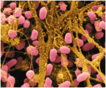
Example: Serratia bacteria are rod-shaped, Gram-negative prokaryotes and opportunistic human pathogens.
The Size, Shape, and Arrangement of Bacterial Cells
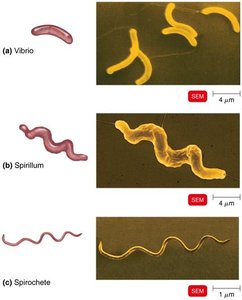
Basic Bacterial Shapes
Bacteria exhibit a variety of shapes and arrangements, which are important for identification and classification.
Bacillus: Rod-shaped
Coccus: Spherical-shaped
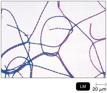
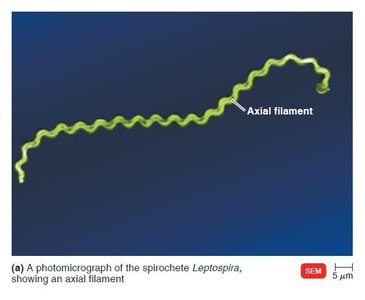
Spiral: Includes vibrio (curved rods), spirillum (helical, rigid), and spirochete (helical, flexible)
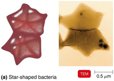
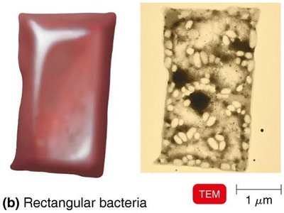
Other shapes: Star-shaped, rectangular
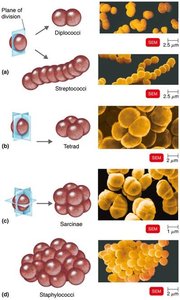
Arrangements of Bacterial Cells
Cocci (spherical):
Diplococci: pairs
Streptococci: chains
Tetrads: groups of four
Sarcinae: cubelike groups of eight
Staphylococci: clusters
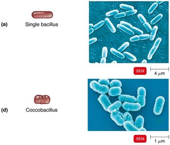
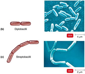
Bacilli (rod-shaped):
Single bacillus
Diplobacilli: pairs
Streptobacilli: chains
Coccobacillus: oval, resembling cocci
Structures External to the Cell Wall
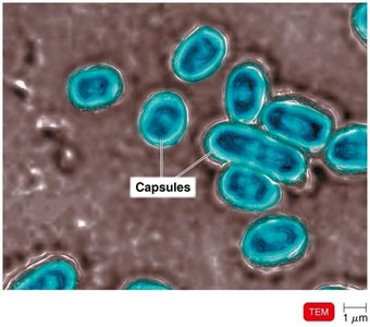
Glycocalyx
The glycocalyx is a viscous, gelatinous polymer external to the cell wall, composed of polysaccharide and/or polypeptide. It exists as either a capsule (organized, firmly attached) or a slime layer (unorganized, loose).
Capsules prevent phagocytosis, contributing to virulence.
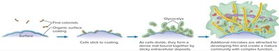
Extracellular polymeric substance (EPS) helps form biofilms, which protect bacteria from antibiotics and immune responses.
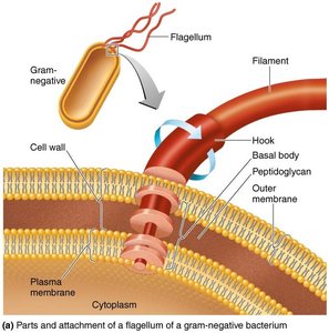
Flagella
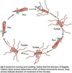
Flagella are long, filamentous appendages that provide motility. They are composed of the protein flagellin and have three main parts: filament, hook, and basal body.
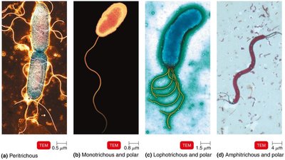
Arrangements include atrichous (none), peritrichous (all over), monotrichous (one at a pole), lophotrichous (tuft at one pole), and amphitrichous (both poles).
Flagella rotate to produce "runs" (straight movement) and "tumbles" (random changes in direction).
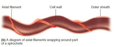
Axial Filaments
Axial filaments (endoflagella) are found in spirochetes. They are anchored at one end and cause the cell to move in a corkscrew motion.
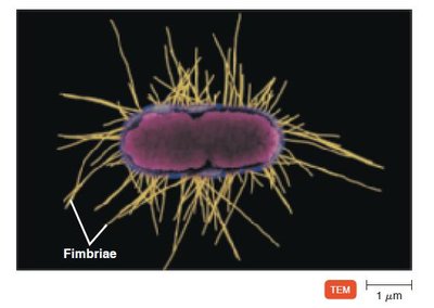
Fimbriae and Pili
Fimbriae: Short, hairlike appendages for attachment; important in biofilm formation.
Pili: Longer; involved in motility (gliding, twitching) and DNA transfer (conjugation pili).
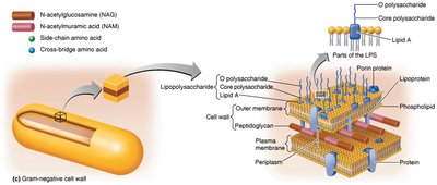
The Cell Wall
Structure and Function
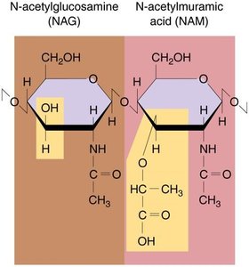
The cell wall prevents osmotic lysis, protects the cell membrane, and contributes to pathogenicity. In bacteria, it is primarily composed of peptidoglycan.
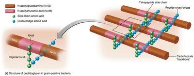
Peptidoglycan is a polymer of alternating N-acetylglucosamine (NAG) and N-acetylmuramic acid (NAM) linked by polypeptides.
Tetrapeptide side chains and peptide cross-bridges provide structural strength.
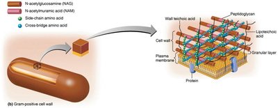
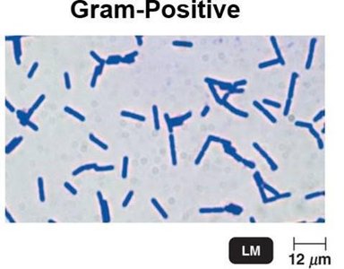
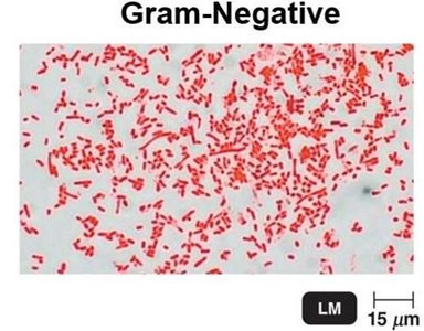
Gram-Positive vs. Gram-Negative Cell Walls
Feature | Gram-Positive | Gram-Negative |
|---|---|---|
Peptidoglycan | Thick | Thin |
Teichoic acids | Present | Absent |
Outer membrane | Absent | Present (LPS) |
Periplasmic space | Absent | Present |
Toxins | Exotoxins | Endotoxins & Exotoxins |
Penicillin susceptibility | High | Low |

Atypical Cell Walls
Acid-fast bacteria: Like gram-positive but with waxy mycolic acid (e.g., Mycobacterium).
Mycoplasmas: Lack cell walls; have sterols in plasma membrane.
Archaea: Cell walls lack peptidoglycan; may have pseudomurein.
Damage to the Cell Wall
Lysozyme: Hydrolyzes bonds in peptidoglycan.
Penicillin: Inhibits peptide bridges in peptidoglycan.
Protoplast: Gram-positive cell with cell wall removed.
Spheroplast: Wall-less gram-negative cell.
Both are susceptible to osmotic lysis.
Structures Internal to the Cell Wall
Plasma (Cytoplasmic) Membrane
The plasma membrane is a phospholipid bilayer with embedded proteins, responsible for selective permeability, ATP production, and, in some bacteria, photosynthesis.
Damage by alcohols, detergents, and antibiotics can cause leakage of cell contents.
Movement Across Membranes
Passive processes: No energy required; includes simple diffusion, facilitated diffusion, and osmosis.
Active processes: Require energy (ATP); includes active transport and group translocation (unique to prokaryotes).
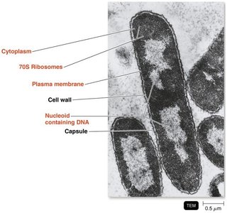
The Nucleoid
The nucleoid contains the bacterial chromosome (circular DNA) and may also contain plasmids (extrachromosomal DNA with non-essential genes).
Endospores
Endospores are highly resistant, dormant structures formed by certain bacteria (e.g., Bacillus, Clostridium) when nutrients are depleted. They resist heat, desiccation, chemicals, and radiation. Sporulation is endospore formation; germination is the return to vegetative state.
Eukaryotic Cells
Flagella and Cilia
Both are projections used for locomotion or moving substances along the cell surface.
Flagella: Long, few in number; Cilia: Short, numerous.
Both consist of microtubules made of tubulin and move in a wavelike manner.
The Cell Wall and Glycocalyx
Cell wall: Found in plants, algae, fungi; made of carbohydrates (cellulose, chitin, glucan).
Glycocalyx: Carbohydrates bonded to proteins and lipids in the plasma membrane; not present in all cells.
The Plasma (Cytoplasmic) Membrane
Similar to prokaryotes: phospholipid bilayer, integral and peripheral proteins.
Differences: Contains sterols and carbohydrates for attachment and recognition.
Functions: Selective permeability, endocytosis (phagocytosis and pinocytosis).
Cytoplasm
Substance inside the plasma membrane and outside the nucleus; 80% water plus proteins, carbs, lipids, ions.
Cytoskeleton provides shape and support.
Ribosomes
Sites of protein synthesis.
Eukaryotic ribosomes: 80S (60S + 40S subunits); membrane-bound or free.
Prokaryotic ribosomes: 70S; found in chloroplasts and mitochondria of eukaryotes.
Organelles
Nucleus: Contains DNA, surrounded by nuclear envelope; DNA complexed with histones as chromatin.
Endoplasmic Reticulum (ER): Rough ER (with ribosomes, protein synthesis); Smooth ER (lipid synthesis).
Golgi Complex: Modifies, sorts, and packages proteins for secretion.
Lysosomes: Contain digestive enzymes.
Vacuoles: Storage and structural support.
Mitochondria: ATP production via cellular respiration.
Chloroplasts: Photosynthesis in plants and algae.
Peroxisomes: Oxidize fatty acids, destroy H2O2.
Centrosomes: Organize microtubules, important in cell division.
The Evolution of Eukaryotes
Endosymbiotic Theory
The endosymbiotic theory proposes that eukaryotic cells evolved when larger prokaryotic cells engulfed smaller ones, which became organelles such as mitochondria and chloroplasts. Evidence includes similarities in DNA, ribosomes, and reproduction between these organelles and prokaryotes.
