 Back
BackFunctional Anatomy of Prokaryotic and Eukaryotic Cells: Structure, Function, and Comparison
Study Guide - Smart Notes
 Tailored notes based on your materials, expanded with key definitions, examples, and context.
Functional Anatomy of Prokaryotic and Eukaryotic Cells
Overview: Prokaryotic vs. Eukaryotic Cells
This section introduces the fundamental differences between prokaryotic and eukaryotic cells, which are the basis for classifying all living organisms. Understanding these differences is essential for microbiology, as it underpins the study of bacteria, archaea, and eukaryotic microbes.
Prokaryotes: Organisms without a true nucleus; DNA is not enclosed by a membrane.
Eukaryotes: Organisms with a true nucleus; DNA is enclosed within a nuclear membrane.
Key Distinctions:
Prokaryotes: Usually one circular chromosome, no histones, no membrane-bound organelles, peptidoglycan (bacteria) or pseudomurein (archaea) cell walls, divide by binary fission.
Eukaryotes: Paired chromosomes in a nuclear membrane, histones present, membrane-bound organelles, polysaccharide cell walls (when present), divide by mitosis.
The Prokaryotic Cell
Size, Shape, and Arrangement of Bacterial Cells
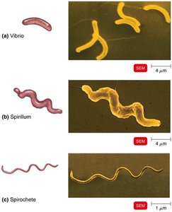
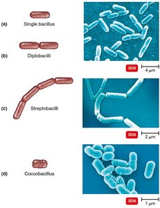
Bacteria exhibit a variety of shapes and arrangements, which are important for identification and classification.
Average Size: 0.2–2.0 μm in diameter, 2–8 μm in length.
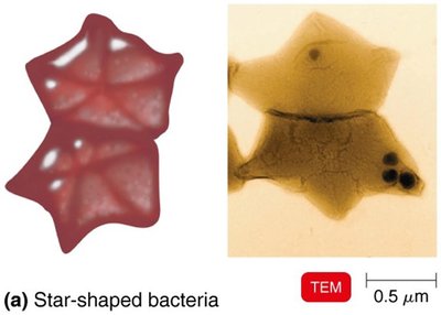
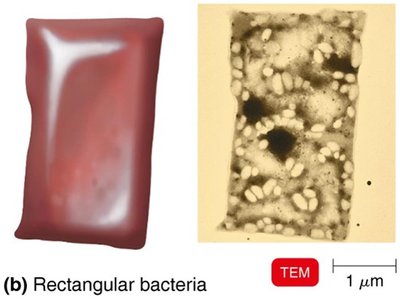
Shapes:
Bacillus: Rod-shaped
Coccus: Spherical-shaped
Spiral: Includes vibrio (curved rods), spirillum (rigid spirals), and spirochete (flexible spirals)
Other: Star-shaped, rectangular
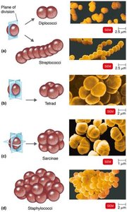
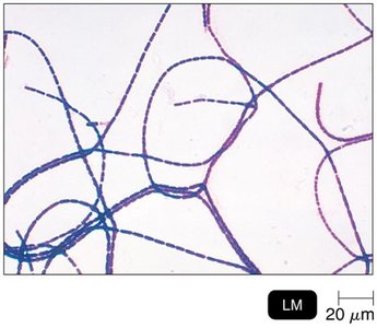
Arrangements:
Pairs: diplococci, diplobacilli
Chains: streptococci, streptobacilli
Clusters: staphylococci
Groups of four: tetrads
Cubelike groups of eight: sarcinae
Structures External to the Cell Wall
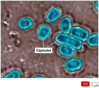
Glycocalyx
The glycocalyx is a gelatinous, sticky substance that surrounds the outside of some bacterial cells. It plays a critical role in pathogenicity and biofilm formation.
Composition: Polysaccharide and/or polypeptide.
Types:
Capsule: Neatly organized and firmly attached.
Slime layer: Unorganized and loosely attached.
Functions:
Contributes to virulence by preventing phagocytosis and aiding in adherence to surfaces (e.g., Bacillus anthracis, Streptococcus pneumoniae).
Extracellular polymeric substance (EPS) helps form biofilms, protecting cells and aiding attachment (e.g., Streptococcus mutans).
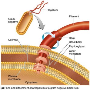
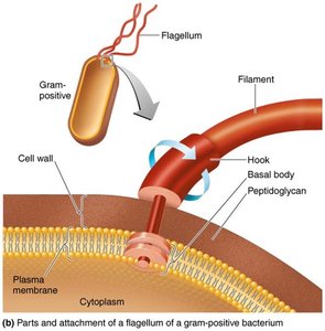
Flagella
Flagella are long, whip-like appendages that provide motility to bacteria. Their structure and arrangement are important for bacterial identification.
Structure: Composed of three parts—filament, hook, and basal body.
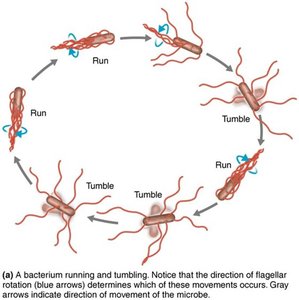
Function: Motility (movement toward or away from stimuli—taxis).
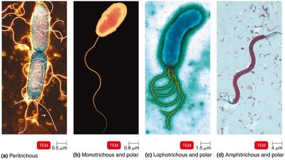
Flagellar Arrangements:
Monotrichous: Single flagellum at one pole
Lophotrichous: Tuft of flagella at one pole
Amphitrichous: Flagella at both poles
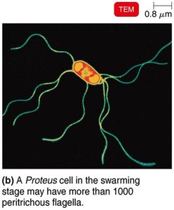
Peritrichous: Flagella distributed over the entire cell
Clinical Relevance: Flagellar proteins (H antigens) are used to distinguish among serovars (e.g., E. coli O157:H7).
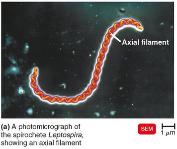
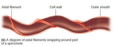
Axial Filaments
Axial filaments, also known as endoflagella, are found in spirochetes and are responsible for their unique corkscrew motility.
Located between the cell wall and outer sheath.
Rotation causes the cell to move in a corkscrew motion.
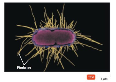
Fimbriae and Pili
Fimbriae and pili are hairlike appendages found on many Gram-negative bacteria, involved in attachment and genetic exchange.
Fimbriae: Allow attachment to surfaces and are important in biofilm formation (e.g., Neisseria gonorrhoeae).
Pili: Involved in motility (gliding, twitching) and conjugation (DNA transfer between cells).
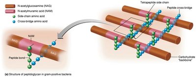
The Cell Wall
Composition and Function
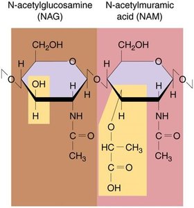
The bacterial cell wall is a rigid structure that provides shape, protection, and is a key target for antibiotics.
Prevents osmotic lysis and protects the cell membrane.
Made of peptidoglycan (in bacteria): a polymer of N-acetylglucosamine (NAG) and N-acetylmuramic acid (NAM) linked by polypeptides.
Penicillin interferes with peptide cross-bridge formation, weakening the cell wall.
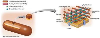
Gram-Positive vs. Gram-Negative Cell Walls
Gram staining differentiates bacteria based on cell wall structure, which has important implications for antibiotic susceptibility and pathogenicity.
Gram-Positive:
Thick peptidoglycan layer
Teichoic acids (lipoteichoic and wall teichoic acids)
High susceptibility to penicillin
Disrupted by lysozyme
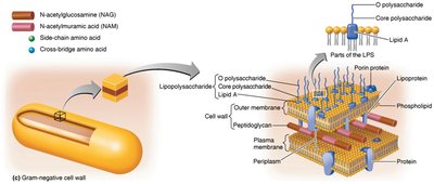
Gram-Negative:
Thin peptidoglycan layer
Outer membrane with lipopolysaccharide (LPS), lipoproteins, and phospholipids
Lipid A (endotoxin), O polysaccharide (antigenic)
Low susceptibility to penicillin
Gram Stain Mechanism
Crystal violet-iodine complexes are retained in Gram-positive cells due to thick peptidoglycan.
Alcohol dissolves the outer membrane of Gram-negative cells, allowing the dye to wash out; cells are counterstained with safranin.
Atypical Cell Walls
Acid-fast bacteria: Thick peptidoglycan with mycolic acid (e.g., Mycobacterium).
Mycoplasmas: Lack cell walls; have sterols in the plasma membrane.
Archaea: May lack cell walls or have walls of pseudomurein (lack NAM and D-amino acids).
Structures Internal to the Cell Wall
Plasma (Cytoplasmic) Membrane
The plasma membrane is a selectively permeable barrier composed of a phospholipid bilayer with embedded proteins.
Functions: Selective permeability, ATP production, photosynthetic pigments (chromatophores in some bacteria).
Fluid Mosaic Model: Membrane components move freely; membrane is self-sealing.
Movement of Materials Across Membranes
Passive Processes:
Simple diffusion: Movement from high to low concentration.
Facilitated diffusion: Transport via membrane proteins.
Osmosis: Movement of water across a selectively permeable membrane.
Active Processes:
Active transport: Requires energy (ATP) to move substances against the gradient.
Group translocation: Substance is chemically modified during transport (requires PEP).
Cytoplasm, Nucleoid, and Ribosomes
Cytoplasm: 80% water, contains DNA, ribosomes, inclusions, and cytoskeleton.
Nucleoid: Region containing the bacterial chromosome (circular, double-stranded DNA) and sometimes plasmids (extrachromosomal DNA).
Ribosomes: Sites of protein synthesis; 70S (50S + 30S subunits); target for antibiotics like streptomycin and erythromycin.
Inclusions and Endospores
Inclusions: Reserve deposits (e.g., phosphate, glycogen, sulfur, gas vacuoles, magnetosomes).
Endospores: Dormant, highly resistant structures formed by Bacillus and Clostridium species under nutrient limitation; survive extreme conditions.
Eukaryotic Cell Structure
Flagella and Cilia
Both are used for movement; flagella are longer and fewer, cilia are shorter and more numerous.
Composed of microtubules in a 9+2 arrangement; move in a wavelike manner.
Cell Wall and Glycocalyx
Cell Wall: Found in plants (cellulose), fungi (chitin), and some protists (glucan, mannan).
Glycocalyx: Carbohydrate-rich layer found in animal cells; involved in cell recognition and attachment.
Plasma Membrane
Similar to prokaryotes but contains sterols and carbohydrates.
Capable of endocytosis (phagocytosis, pinocytosis, receptor-mediated).
Cytoplasm and Ribosomes
Cytoskeleton composed of microfilaments, intermediate filaments, and microtubules.
80S ribosomes (60S + 40S subunits) in cytoplasm and on rough ER; 70S ribosomes in mitochondria and chloroplasts.
Organelles
Nucleus: Contains DNA complexed with histones; surrounded by a double membrane with nuclear pores.
Endoplasmic Reticulum (ER): Rough ER (with ribosomes, protein synthesis), Smooth ER (lipid synthesis).
Golgi Complex: Modifies, sorts, and packages proteins and lipids.
Lysosomes: Contain digestive enzymes.
Vacuoles: Storage and structural support.
Mitochondria: Site of ATP production; contain their own DNA and 70S ribosomes.
Chloroplasts: Site of photosynthesis in plants and algae; contain their own DNA and 70S ribosomes.
Peroxisomes: Oxidize fatty acids and destroy hydrogen peroxide.
Centrosomes: Organize microtubules and are important in cell division.
Endosymbiotic Theory
The endosymbiotic theory explains the origin of mitochondria and chloroplasts as descendants of free-living bacteria engulfed by ancestral eukaryotic cells.
Evidence: Mitochondria and chloroplasts resemble bacteria in size and shape, have circular DNA, reproduce independently, and contain 70S ribosomes.
Additional info: This summary covers the main structural and functional features of prokaryotic and eukaryotic cells, with emphasis on their differences, specialized structures, and clinical relevance. It is suitable for exam preparation in a college-level microbiology course.
