 Back
BackFunctional Anatomy of Prokaryotic and Eukaryotic Cells: Structure, Function, and Diversity
Study Guide - Smart Notes
 Tailored notes based on your materials, expanded with key definitions, examples, and context.
Functional Anatomy of Prokaryotic and Eukaryotic Cells
Overview: Prokaryotic vs. Eukaryotic Cells
This section introduces the fundamental differences between prokaryotic and eukaryotic cells, which are the two primary cell types in microbiology. Understanding these differences is essential for classifying microorganisms and predicting their biological behaviors.
Prokaryotes: Cells lacking a true nucleus and membrane-bound organelles. Includes Bacteria and Archaea.
Eukaryotes: Cells with a true nucleus enclosed by a nuclear membrane and various membrane-bound organelles. Includes fungi, algae, protozoa, and helminths.
Key Differences:
Prokaryotes: Usually one circular chromosome (not in a membrane), no histones, no membrane-enclosed organelles, cell walls (peptidoglycan in bacteria, pseudomurein in archaea), divide by binary fission.
Eukaryotes: Paired chromosomes in a nuclear membrane, histones present, organelles present, polysaccharide cell walls (when present), divide by mitosis.
The Prokaryotic Cell
Size, Shape, and Arrangement of Bacterial Cells
Bacteria exhibit a variety of shapes and arrangements, which are important for identification and classification.
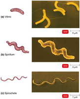
Shapes:
Bacillus: Rod-shaped
Coccus: Spherical-shaped
Spiral forms: Vibrio (curved rods), Spirillum (rigid spirals), Spirochete (flexible spirals)
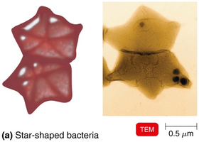
Other shapes: Star-shaped, rectangular
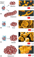
Arrangements:
Pairs: diplococci, diplobacilli
Chains: streptococci, streptobacilli
Clusters: staphylococci
Groups of four: tetrads
Cubelike groups of eight: sarcinae
Structure of a Prokaryotic Cell
Prokaryotic cells have a simple structure but possess specialized features that contribute to their survival and pathogenicity.
Major components: Cell wall, plasma membrane, cytoplasm, nucleoid, ribosomes, inclusions, and sometimes external structures (glycocalyx, flagella, fimbriae, pili).
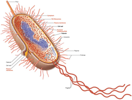
Structures External to the Cell Wall
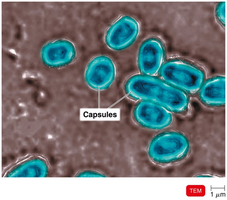
Glycocalyx
The glycocalyx is a viscous, gelatinous polymer external to the cell wall, composed of polysaccharide and/or polypeptide. It exists as either a capsule (organized, firmly attached) or a slime layer (unorganized, loose).
Functions:
Contributes to virulence by preventing phagocytosis and aiding in adherence to surfaces (e.g., Bacillus anthracis, Streptococcus pneumoniae).
Facilitates biofilm formation, protecting cells and aiding attachment (e.g., Streptococcus mutans, Vibrio cholerae).
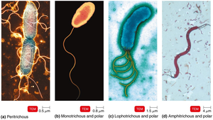
Flagella
Flagella are long, filamentous appendages that provide motility to bacteria. They are composed of the protein flagellin and consist of three parts: filament, hook, and basal body.
Function: Motility (movement toward or away from stimuli—taxis), identification (H antigens).
Arrangement types: Peritrichous (all over), monotrichous (single, polar), lophotrichous (tuft at one pole), amphitrichous (both poles).
Axial Filaments and Archaella
Axial filaments (endoflagella): Found in spirochetes, anchored at one end, rotation causes corkscrew movement.
Archaella: Motility structures in Archaea, made of archaellins, rotate like flagella, use ATP for energy.
Fimbriae and Pili
Fimbriae and pili are hairlike appendages found on many Gram-negative bacteria.
Fimbriae: Enable attachment to surfaces and biofilm formation (e.g., Neisseria gonorrhoeae, E. coli O157).
Pili: Involved in motility (gliding, twitching) and DNA transfer (conjugation pili).

The Cell Wall
Composition and Function
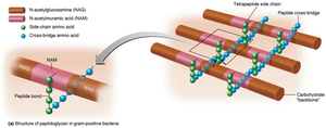
The bacterial cell wall is a rigid structure that protects the cell, maintains its shape, and prevents osmotic lysis. It is a major target for antibiotics and a key factor in pathogenicity.
Peptidoglycan: Main component in bacteria; a polymer of N-acetylglucosamine (NAG) and N-acetylmuramic acid (NAM) linked by polypeptides, forming a lattice structure.
Penicillin: Inhibits peptide cross-bridge formation, weakening the cell wall.
Gram-Positive vs. Gram-Negative Cell Walls
Gram-Positive:
Thick peptidoglycan layer, teichoic acids (lipoteichoic and wall teichoic acids), two rings in flagellar basal body, high susceptibility to penicillin, disrupted by lysozyme, produce exotoxins.
Gram-Negative:
Thin peptidoglycan, outer membrane (lipopolysaccharide, lipoproteins, phospholipids), periplasmic space, four rings in flagellar basal body, low susceptibility to penicillin, produce endotoxins and exotoxins, protected from phagocytes and antibiotics.
Atypical Cell Walls
Acid-fast bacteria: Thick peptidoglycan, waxy mycolic acid, stain with carbolfuchsin (e.g., Mycobacterium, Nocardia).
Mycoplasmas: Lack cell walls, sterols in plasma membrane.
Archaea: Wall-less or walls of pseudomurein (lack NAM and D-amino acids).
Damage to the Cell Wall
Lysozyme: Hydrolyzes bonds in peptidoglycan, especially effective against Gram-positive bacteria.
Penicillin: Inhibits peptide bridge formation in peptidoglycan.
Protoplast: Wall-less Gram-positive cell.
Spheroplast: Wall-less Gram-negative cell.
L forms: Wall-less cells that swell into irregular shapes; all are susceptible to osmotic lysis.
Structures Internal to the Cell Wall
Plasma (Cytoplasmic) Membrane
The plasma membrane is a phospholipid bilayer with embedded proteins, responsible for selective permeability, ATP production, and sometimes photosynthesis (chromatophores).
Fluid mosaic model: Membrane is dynamic, proteins and lipids move laterally.
Glycoproteins and glycolipids: Some membrane components have attached carbohydrates.
Movement of Materials Across Membranes
Passive processes: No energy required; substances move from high to low concentration.
Simple diffusion: Movement of solute until equilibrium is reached.
Facilitated diffusion: Transport via membrane proteins (channels or carriers).
Osmosis: Net movement of water across a selectively permeable membrane.
Active processes: Require energy (ATP); substances move against the concentration gradient.
Active transport: Transporter proteins and ATP move ions, amino acids, sugars.
Cytoplasm and Internal Structures
Cytoplasm: Thick, aqueous solution containing water, proteins, carbohydrates, lipids, ions, DNA, ribosomes, inclusions.
Cytoskeleton: Fibers involved in cell division, shape, growth, and DNA movement.
Nucleoid
Bacterial chromosome: Circular, double-stranded DNA, not membrane-bound, no histones.
Plasmids: Small, extrachromosomal DNA circles; carry nonessential genes (e.g., antibiotic resistance), replicate independently, can be transferred between cells.
Ribosomes
Function: Protein synthesis.
Structure: Composed of protein and rRNA; 70S in prokaryotes.
Antibiotic targets: Streptomycin, gentamicin, erythromycin, chloramphenicol inhibit prokaryotic ribosomes.
Inclusions
Function: Reserve deposits of nutrients.
Types:
Metachromatic granules (phosphate reserves)
Polysaccharide granules (energy reserves)
Lipid inclusions (energy reserves)
Sulfur granules (energy reserves)
Carboxysomes (contain RuBisCO for CO2 fixation)
Gas vacuoles (buoyancy)
Magnetosomes (iron oxide, destroy H2O2)
Endospores
Endospores are highly resistant, dormant structures formed by certain bacteria (e.g., Bacillus, Clostridium) when nutrients are depleted. They can survive extreme conditions and are important in food safety and sterilization.
Sporulation: Formation of endospores.
Germination: Return to vegetative state when conditions improve.
Additional info: This guide covers the essential structural and functional features of prokaryotic cells, with emphasis on their diversity, adaptations, and relevance to microbiology. The included images illustrate key bacterial shapes, arrangements, and structural features, directly supporting the textual explanations.
