Back
BackFunctional Anatomy of Prokaryotic and Eukaryotic Cells: Microbiology Study Notes
Study Guide - Smart Notes
 Tailored notes based on your materials, expanded with key definitions, examples, and context.
Functional Anatomy of Prokaryotic and Eukaryotic Cells
Overview: Prokaryotic vs. Eukaryotic Cells
This section compares the fundamental differences between prokaryotic and eukaryotic cells, which are the two primary cell types in microbiology. Understanding these differences is essential for classifying microorganisms and understanding their biology.
Prokaryotes: One circular chromosome (not in a membrane), no histones, no organelles, cell walls (peptidoglycan in Bacteria, pseudomurein in Archaea), divide by binary fission.
Eukaryotes: Paired chromosomes in a nuclear membrane, histones present, organelles present, cell walls (when present) made of polysaccharides, divide by mitosis.
Shapes and Arrangements of Prokaryotic Cells
Prokaryotic cells exhibit a variety of shapes and arrangements, which are important for identification and classification.
Common shapes: Bacillus (rod-shaped), Coccus (spherical), Spiral (twisted or curved).
Exceptional shapes: Star-shaped, Rectangular.
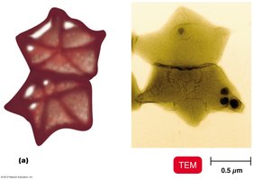
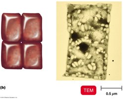
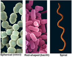
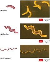
Spiral forms: Vibrio (comma-shaped), Spirillum (rigid spiral), Spirochete (flexible spiral).
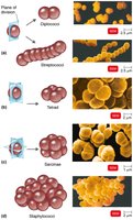
Arrangements: Diplococci/diplobacilli (pairs), Streptococci/streptobacilli (chains), Staphylococci (clusters), Tetrads (groups of four), Sarcinae (cubical groups of eight).
Structure of a Prokaryotic Cell
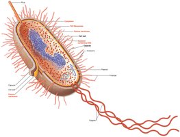
Prokaryotic cells have a complex structure with specialized components that contribute to their survival and pathogenicity.
Glycocalyx
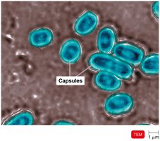
The glycocalyx is a viscous, gelatinous layer external to the cell wall, composed of polysaccharide and/or polypeptide. It exists as either a capsule (organized, firmly attached) or a slime layer (unorganized, loose).
Functions: Contributes to virulence by preventing phagocytosis and aiding in biofilm formation.
Flagella
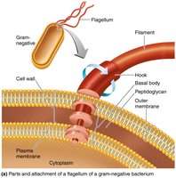
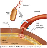
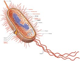
Flagella are filamentous appendages that provide motility to bacteria. They are composed of the protein flagellin and consist of three parts: filament, hook, and basal body.
Function: Propel bacteria through liquid environments.
Arrangements of Bacterial Flagella
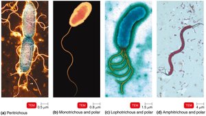
Peritrichous: Flagella distributed over the entire cell.
Monotrichous: Single flagellum at one pole.
Lophotrichous: Tuft of flagella at one pole.
Amphitrichous: Flagella at both poles.
Fimbriae and Pili
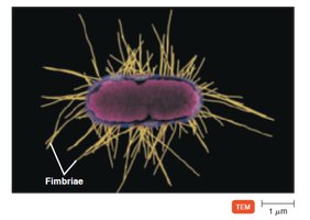
Fimbriae are hairlike appendages that allow for attachment to surfaces, while pili are involved in motility and DNA transfer (conjugation).
The Cell Wall
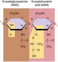
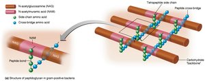
The bacterial cell wall provides structural support, prevents osmotic lysis, and contributes to pathogenicity. It is primarily composed of peptidoglycan, a polymer of N-acetylglucosamine (NAG) and N-acetylmuramic acid (NAM) linked by polypeptides.
Gram-Positive vs. Gram-Negative Cell Walls
Feature | Gram-Positive | Gram-Negative |
|---|---|---|
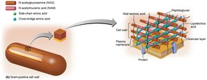
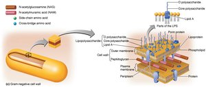
Peptidoglycan | Thick | Thin |
Teichoic acids | Present | Absent |
Outer membrane | Absent | Present (lipopolysaccharides, lipoproteins, phospholipids) |
Periplasmic space | Absent | Present |
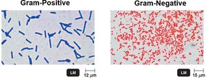
Gram Stain Mechanism
The Gram stain differentiates bacteria based on cell wall structure:
Gram-positive: Alcohol dehydrates peptidoglycan, trapping crystal violet-iodine complexes.
Gram-negative: Alcohol dissolves outer membrane, allowing dye to wash out; cells are counterstained with safranin.
Damage to the Cell Wall
Lysozyme: Hydrolyzes bonds in peptidoglycan.
Penicillin: Inhibits peptide bridge formation in peptidoglycan.
Protoplast: Wall-less gram-positive cell.
Spheroplast: Wall-less gram-negative cell.
L forms: Wall-less cells that swell into irregular shapes.
The Plasma (Cytoplasmic) Membrane
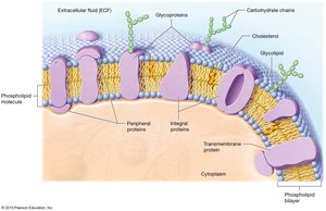
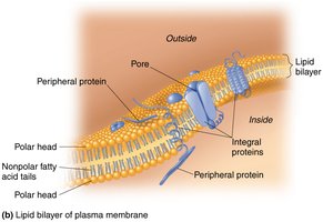
The plasma membrane is a phospholipid bilayer with embedded proteins, responsible for selective permeability and metabolic functions.
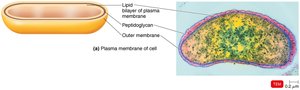
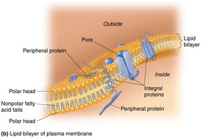
Functions: Selective permeability, ATP production, photosynthetic pigments (chromatophores in some bacteria).
Transport Across the Membrane
Simple diffusion: Movement of solute from high to low concentration until equilibrium is reached.
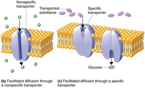
Facilitated diffusion: Solute combines with transporter protein; moves ions and larger molecules with the gradient.
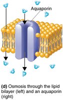
Osmosis: Movement of water across a selectively permeable membrane from high to low water concentration, via lipid layer or aquaporins.
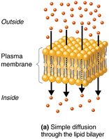
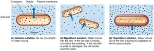
Osmotic conditions: Isotonic (no net water movement), Hypotonic (water enters cell), Hypertonic (water leaves cell).
Active transport: Requires transporter protein and ATP; moves substances against the gradient.
Cytoplasm and Internal Structures
The cytoplasm is the substance inside the plasma membrane, containing water, proteins, carbohydrates, lipids, ions, and a cytoskeleton.
Nucleoid: Contains the bacterial chromosome (circular DNA) and plasmids (extrachromosomal DNA).
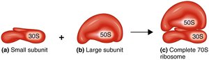
Ribosomes: Sites of protein synthesis; 70S (50S + 30S subunits) in prokaryotes.
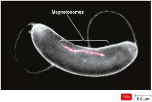
Inclusions: Reserve deposits (e.g., metachromatic granules, polysaccharide granules, lipid inclusions, sulfur granules, carboxysomes, gas vacuoles, magnetosomes).
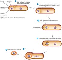
Endospores: Resting cells formed under nutrient depletion; highly resistant to environmental stress. Produced by Bacillus and Clostridium. Sporulation is endospore formation; germination is return to vegetative state.
Eukaryotic Cell Structure
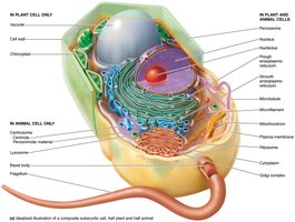
Overview of Eukaryotic Cell Structures
Eukaryotic cells are structurally more complex than prokaryotic cells, containing membrane-bound organelles and a defined nucleus.
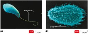
Flagella and Cilia
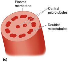
Flagella and cilia are projections used for locomotion or moving substances along the cell surface. Both are composed of microtubules arranged in a 9+2 pattern.
Flagella: Long, few in number; move in a wavelike manner.
Cilia: Short, numerous; move substances along the cell surface.
The Cell Wall and Glycocalyx in Eukaryotes
Cell wall: Found in plants, algae, fungi; composed of carbohydrates (cellulose, chitin, glucan, mannan).
Glycocalyx: Carbohydrates bonded to proteins and lipids in the plasma membrane; found in animal cells.
The Plasma (Cytoplasmic) Membrane in Eukaryotes
The eukaryotic plasma membrane is similar to that of prokaryotes but contains sterols and carbohydrates for attachment and recognition. It is involved in selective permeability and transport processes, including endocytosis (phagocytosis and pinocytosis).
Cytoplasm and Cytoskeleton
Cytoplasm: Substance inside the plasma membrane and outside the nucleus.
Cytosol: Fluid portion of cytoplasm.
Cytoskeleton: Microfilaments and intermediate filaments provide shape and support.
Cytoplasmic streaming: Movement of cytoplasm throughout the cell.
Ribosomes
80S ribosomes: Large (60S) and small (40S) subunits; membrane-bound (attached to ER) or free in cytoplasm.
70S ribosomes: Found in chloroplasts and mitochondria.
The Nucleus
The nucleus is a double-membrane structure containing the cell’s DNA, complexed with histones to form chromatin. During cell division, chromatin condenses into chromosomes.
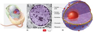
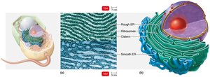
Endomembrane System: ER and Golgi Complex
Endoplasmic Reticulum (ER): Folded transport network; rough ER (with ribosomes) synthesizes proteins, smooth ER synthesizes membranes, fats, and hormones.
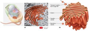
Golgi Complex: Modifies and transports proteins from the ER via secretory vesicles to the plasma membrane.
Other Eukaryotic Organelles
Lysosomes: Contain digestive enzymes; formed in the Golgi complex.
Vacuoles: Storage and shape; bring food into cells.
Peroxisomes: Oxidize fatty acids; destroy hydrogen peroxide (H2O2).
Centrosomes: Form the mitotic spindle; critical for cell division.
Mitochondria
Mitochondria are double-membraned organelles with inner folds (cristae) and matrix, responsible for ATP production via cellular respiration.
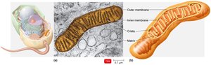
Chloroplasts
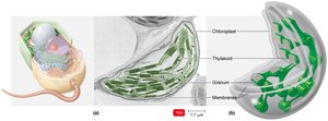
Chloroplasts are the sites of photosynthesis in plant and algal cells, containing thylakoids with chlorophyll.