 Back
Back(Chapter 4) Functional Anatomy of Prokaryotic and Eukaryotic Cells: Study Notes
Study Guide - Smart Notes
 Tailored notes based on your materials, expanded with key definitions, examples, and context.
Functional Anatomy of Prokaryotic and Eukaryotic Cells
Comparing Prokaryotic and Eukaryotic Cells: An Overview
Prokaryotic and eukaryotic cells differ fundamentally in their structure and organization. Understanding these differences is essential for microbiology students, as it forms the basis for classifying microorganisms and understanding their biology.
Prokaryote: Derived from Greek for 'prenucleus'. Characterized by a single circular chromosome not enclosed in a membrane, absence of histones, and lack of membrane-bound organelles. Bacteria have peptidoglycan cell walls, while Archaea have pseudomurein cell walls. Division occurs by binary fission.
Eukaryote: Derived from Greek for 'true nucleus'. Possess paired chromosomes within a nuclear membrane, histones, and membrane-bound organelles. Cell walls, when present, are made of polysaccharides. Division occurs by mitosis.
The Size, Shape, and Arrangement of Bacterial Cells
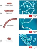
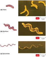
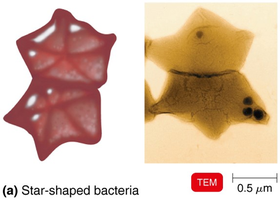
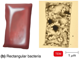
Bacteria exhibit a variety of shapes and arrangements, which are important for identification and classification. Most bacteria are monomorphic (single shape), but some are pleomorphic (variable shapes).
Common shapes: Bacillus (rod-shaped), Coccus (spherical), Spiral (including Vibrio, Spirillum, Spirochete), Star-shaped, and Rectangular.
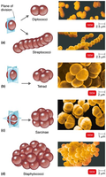
Arrangements: Pairs (diplococci, diplobacilli), Clusters (staphylococci), Chains (streptococci, streptobacilli), Groups of four (tetrads), Cubelike groups of eight (sarcinae).
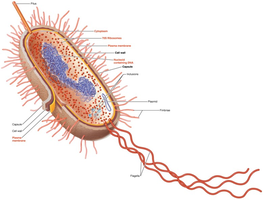
The Structure of a Prokaryotic Cell
Prokaryotic cells have a simple structure but contain specialized components that enable their survival and function.
Cell wall: Provides structural support and protection.
Plasma membrane: Controls the movement of substances in and out of the cell.
Cytoplasm: Contains DNA, ribosomes, and inclusions.
External structures: Include glycocalyx, flagella, fimbriae, and pili.
Glycocalyx
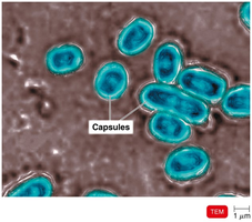
The glycocalyx is an external layer that can be either a capsule or a slime layer. It plays a crucial role in virulence and biofilm formation.
Capsule: Neatly organized and firmly attached; prevents phagocytosis and aids in adherence.
Slime layer: Unorganized and loose; helps in biofilm formation.
Function: Protects cells, helps microbes attach to surfaces, and contributes to pathogenicity.
Flagella
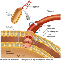
Flagella are filamentous appendages that provide motility to bacteria. They consist of three parts: filament, hook, and basal body.
Function: Allow bacteria to move toward or away from stimuli (taxis).
Structure: Filament (outermost), hook (attachment), basal body (anchors to cell wall and membrane).
Flagella proteins: Serve as H antigens for serovar identification.
Fimbriae and Pili
Fimbriae and pili are hairlike appendages found on prokaryotic cells.
Fimbriae: Enable attachment to surfaces and are involved in biofilm formation.
Pili: Involved in motility (gliding and twitching) and DNA transfer (conjugation pili).
The Cell Wall
The cell wall is a critical structure that prevents osmotic lysis and contributes to pathogenicity. Its chemical composition is used to differentiate major groups of bacteria.
Peptidoglycan: Polymer of N-acetylglucosamine (NAG) and N-acetylmuramic acid (NAM) linked by polypeptides.
Penicillin: Interferes with peptide cross-bridges, weakening the cell wall.
Gram-Positive Cell Walls
Gram-positive bacteria have thick peptidoglycan layers and teichoic acids.
Teichoic acids: Provide antigenic specificity and regulate cation movement.
Lipoteichoic acid: Links cell wall to plasma membrane.
Gram-Negative Cell Walls
Gram-negative bacteria have thin peptidoglycan, an outer membrane, and a periplasmic space.
Outer membrane: Contains lipopolysaccharide (LPS), lipoproteins, and phospholipids.
LPS: O polysaccharide (antigen), Lipid A (endotoxin).
Porins: Form channels through the membrane.
Cell Walls and the Gram Stain Mechanism
The Gram stain differentiates bacteria based on cell wall structure.
Gram-positive: Alcohol dehydrates peptidoglycan, retaining crystal violet-iodine complex.
Gram-negative: Alcohol dissolves outer membrane, washes out dye, cells are colorless until counterstained with safranin.
Atypical Cell Walls
Some bacteria have atypical cell walls, such as acid-fast cell walls.
Acid-fast cell walls: Thick peptidoglycan with waxy mycolic acid; stain with carbolfuchsin.
Genera: Mycobacterium, Nocardia.
Damage to the Cell Wall
Certain agents can damage the bacterial cell wall, making cells susceptible to osmotic lysis.
Lysozyme: Hydrolyzes bonds in peptidoglycan.
Penicillin: Inhibits peptide bridge formation.
Protoplast: Wall-less gram-positive cell.
Spheroplast: Wall-less gram-negative cell.
L forms: Wall-less cells that swell into irregular shapes.
The Plasma (Cytoplasmic) Membrane Structure
The plasma membrane is a phospholipid bilayer with embedded proteins and carbohydrates.
Fluid mosaic model: Membrane is flexible; proteins and phospholipids move freely.
Glycoproteins and glycolipids: Carbohydrate-attached membrane components.
The Movement of Materials across Membranes
Substances move across membranes by passive or active processes.
Passive processes: No energy required; substances move from high to low concentration.
Active processes: Energy required; substances move from low to high concentration.
Passive Processes
Simple diffusion: Movement of solute from high to low concentration until equilibrium is reached.
Facilitated diffusion: Uses integral membrane proteins as channels or carriers.
Osmosis: Net movement of water across a selectively permeable membrane.
Osmotic pressure: Pressure needed to stop water movement.
Isotonic solution: Equal solute concentrations; no net water movement.
Hypotonic solution: Lower solute outside; water moves into cell.
Hypertonic solution: Higher solute outside; water moves out of cell.
Active Processes
Active transport: Requires transporter protein and ATP; moves substances against gradient.
Group translocation: Requires transporter protein and PEP; substance is chemically altered during transport.
Cytoplasm
The cytoplasm is a thick, aqueous substance inside the plasma membrane, containing DNA, ribosomes, and inclusions.
Cytoskeleton: Fibers involved in cell division, shape, growth, and DNA movement.
The Nucleoid
The nucleoid contains the bacterial chromosome and plasmids.
Bacterial chromosome: Circular, double-stranded DNA; not membrane-bound.
Plasmids: Small, extrachromosomal DNA circles; carry nonessential genes (e.g., antibiotic resistance).
Ribosomes
Ribosomes are the sites of protein synthesis.
Structure: Made of protein and ribosomal RNA; 70S in prokaryotes.
Antibiotics: Streptomycin, gentamicin, erythromycin, and chloramphenicol target prokaryotic ribosomes.
Inclusions
Inclusions are reserve deposits of nutrients and other substances.
Types: Metachromatic granules (phosphate), polysaccharide granules (energy), lipid inclusions (energy), sulfur granules (energy), carboxysomes (RuBisCO enzyme), gas vacuoles (buoyancy), magnetosomes (iron oxide).
Endospores
Endospores are highly resistant, dormant structures formed by certain bacteria when nutrients are depleted.
Resistant to: Desiccation, heat, chemicals, radiation.
Produced by: Bacillus and Clostridium.
Sporulation: Endospore formation.
Germination: Return to vegetative state.
Importance: Survival mechanism; significant in food industry.
Additional info: These notes expand on brief points with academic context, definitions, and examples to ensure completeness and clarity for exam preparation.
