 Back
BackFunctional Anatomy of Prokaryotic and Eukaryotic Cells: Microbiology Study Notes
Study Guide - Smart Notes
 Tailored notes based on your materials, expanded with key definitions, examples, and context.
Comparing Prokaryotic and Eukaryotic Cells
Cell Structure Overview
Prokaryotic and eukaryotic cells differ fundamentally in their structural organization, genetic material, and cellular processes. Understanding these differences is essential for microbiology students.
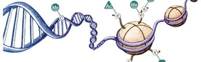
Prokaryotes: Characterized by the absence of a membrane-bound nucleus and organelles. Their genetic material is a single circular chromosome located in the nucleoid.
Eukaryotes: Possess a true nucleus surrounded by a nuclear membrane and various membrane-bound organelles. Their genetic material is organized into paired chromosomes.
Feature | Prokaryote | Eukaryote |
|---|---|---|
Nucleus | No (nucleoid) | Yes (membrane-bound) |
Chromosomes | One circular | Paired, linear |
Organelles | Absent | Present |
Cell Wall | Peptidoglycan (Bacteria), Pseudomurein (Archaea) | Polysaccharides (plants, fungi, some protists) |
Division | Binary fission | Mitosis |
The Size, Shape, and Arrangement of Bacterial Cells
Bacterial Morphology
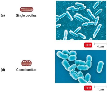
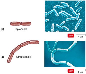
Bacteria exhibit a variety of shapes and arrangements, which are important for identification and classification.
Average size: 0.2–2.0 μm diameter, 2–8 μm length
Monomorphic: Most bacteria have a single, consistent shape
Pleomorphic: Some bacteria can vary in shape
Basic Shapes
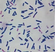
Bacillus: Rod-shaped
Coccus: Spherical-shaped
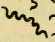
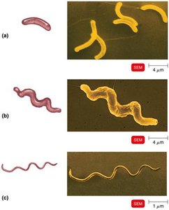
Spiral: Includes vibrio (comma-shaped), spirillum (rigid), and spirochete (flexible)
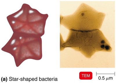
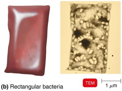
Star-shaped and Rectangular: Rare morphologies
Arrangements
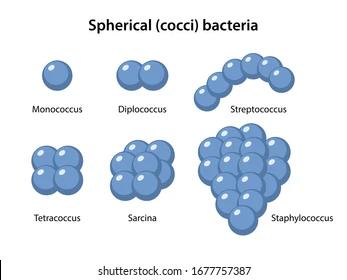
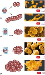
Pairs: Diplococci, diplobacilli
Chains: Streptococci, streptobacilli
Clusters: Staphylococci
Groups of four: Tetrads
Cubelike groups of eight: Sarcinae
Structure of a Prokaryotic Cell
Cell Components
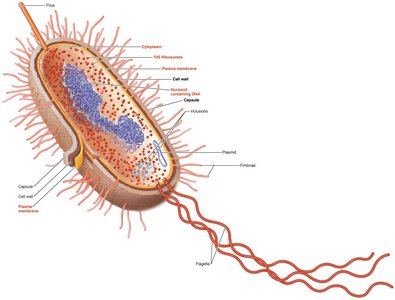
Prokaryotic cells contain several key structures, each with specific functions.
Capsule: External polysaccharide layer contributing to virulence
Cell wall: Provides shape and protection
Plasma membrane: Selective permeability and metabolic functions
Cytoplasm: Contains ribosomes, nucleoid, and inclusions
Flagella, fimbriae, pili: Appendages for movement and attachment
Structures External to the Cell Wall
Glycocalyx
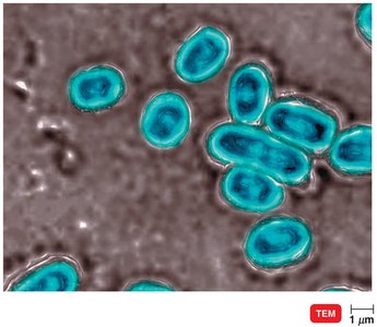
The glycocalyx is a viscous, gelatinous layer external to the cell wall, composed of polysaccharides and/or polypeptides.
Capsule: Organized, firmly attached; prevents phagocytosis
Slime layer: Unorganized, loosely attached
Biofilms: Glycocalyx helps bacteria adhere to surfaces and form biofilms
Flagella
Flagella are filamentous appendages used for motility.
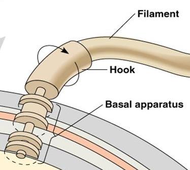
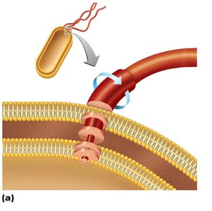
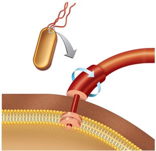
Structure: Composed of filament, hook, and basal body
Basal body: Anchors flagellum; arrangement differs in gram-positive (2 rings) and gram-negative (4 rings) bacteria
Function: Enables movement via "run" and "tumble"; taxis (movement toward/away from stimuli)
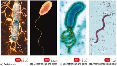
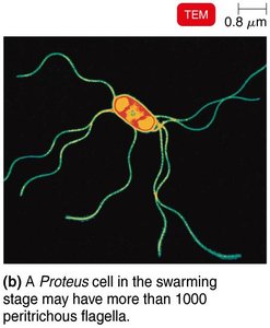
Flagella Arrangements
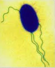
Monotrichous: Single flagellum at one end
Lophotrichous: Tuft of flagella at one end
Amphitrichous: Flagella at both ends
Peritrichous: Flagella all over the cell
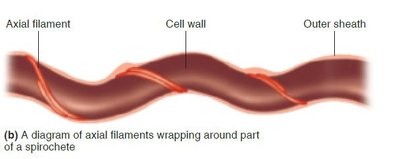
Axial Filaments
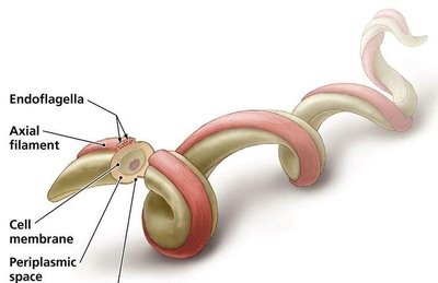
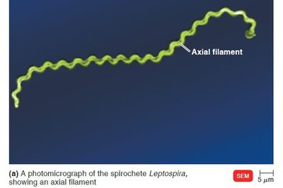
Axial filaments, or endoflagella, are found in spirochetes and enable corkscrew movement.
Structure: Anchored at one end, wraps around cell
Function: Rotation causes movement
Fimbriae and Pili
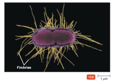
Fimbriae: Hairlike appendages for attachment to surfaces and other cells
Pili: Longer, involved in motility and DNA transfer (conjugation)
The Cell Wall
Composition and Function
The cell wall is a critical structure for bacterial shape, protection, and classification.
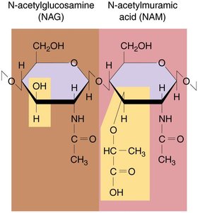
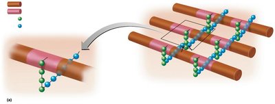
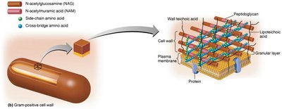
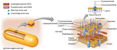
Peptidoglycan: Polymer of N-acetylglucosamine (NAG) and N-acetylmuramic acid (NAM) linked by polypeptides
Function: Prevents osmotic lysis, anchors flagella, site of antibiotic action
Gram-Positive vs. Gram-Negative Cell Walls
Gram-Positive: Thick peptidoglycan, teichoic acids, no outer membrane
Gram-Negative: Thin peptidoglycan, outer membrane with lipopolysaccharides (LPS), periplasmic space
Feature | Gram-Positive | Gram-Negative |
|---|---|---|
Peptidoglycan | Thick | Thin |
Teichoic acids | Present | Absent |
Outer membrane | Absent | Present |
LPS | None | High |
Basal body rings | 2 | 4 |
Toxins | Exotoxins | Endotoxins & Exotoxins |
Antibiotic resistance | Low | High |
Gram Stain Mechanism
The Gram stain differentiates bacteria based on cell wall structure.
Crystal violet: Primary stain
Iodine: Mordant
Alcohol: Decolorizer
Safranin: Counterstain
Gram-positive: Retain purple color
Gram-negative: Stain pink
Atypical Cell Walls
Acid-fast: Waxy mycolic acid layer (e.g., Mycobacterium)
Mycoplasmas: Lack cell walls, contain sterols
Archaea: Wall-less or pseudomurein walls
Structures Internal to the Cell Wall
Plasma Membrane
The plasma membrane is a phospholipid bilayer with embedded proteins, responsible for selective permeability and metabolic functions.
Fluid mosaic model: Proteins and lipids move freely
Damage: Alcohols, detergents, and antibiotics can disrupt membrane integrity
Movement of Materials Across Membranes
Passive processes: Simple diffusion, facilitated diffusion, osmosis
Active processes: Active transport (requires ATP), group translocation (unique to prokaryotes)
Cytoplasm, Nucleoid, Ribosomes, and Inclusions
Cytoplasm: 80% water, contains proteins, carbohydrates, lipids, ions
Nucleoid: Contains bacterial chromosome (DNA)
Plasmids: Extrachromosomal DNA, often carry antibiotic resistance genes
Ribosomes: 70S (prokaryotes), site of protein synthesis
Inclusions: Reserve deposits (e.g., phosphate, polysaccharide, lipid, sulfur granules, gas vacuoles, magnetosomes)
Endospores
Endospores are highly resistant, dormant structures formed by certain bacteria (e.g., Bacillus, Clostridium) under adverse conditions.
Sporulation: Formation of endospore
Germination: Return to vegetative state
Resistant to: Heat, desiccation, chemicals, radiation
Eukaryotic Cell Structures
Organelles and Functions
Eukaryotic cells contain specialized organelles, each with distinct functions.
Nucleus: Contains DNA, surrounded by nuclear envelope
Endoplasmic reticulum: Rough (protein synthesis), smooth (lipid synthesis)
Golgi complex: Modifies and transports proteins
Lysosomes: Digestive enzymes
Vacuoles: Storage and shape
Mitochondria: ATP production
Chloroplasts: Photosynthesis
Peroxisomes: Oxidation of fatty acids
Centrosomes: Cell division
Flagella and Cilia
Flagella: Long, few; movement
Cilia: Short, numerous; movement and substance transport
Structure: 9+2 arrangement of microtubules (tubulin)
Cell Wall and Glycocalyx
Cell wall: Plants (cellulose), fungi (chitin), yeasts (glucan, mannan)
Glycocalyx: Carbohydrate layer for attachment and recognition (animal cells)
Plasma Membrane
Contains sterols: Complex lipids
Contains carbohydrates: Attachment and cell recognition
Endocytosis: Phagocytosis and pinocytosis (not in prokaryotes)
Cytoplasm and Ribosomes
Cytoskeleton: Microfilaments and intermediate filaments
Cytoplasmic streaming: Movement of cytoplasm (eukaryotes only)
Ribosomes: 80S (cytoplasm), 70S (mitochondria and chloroplasts)
The Evolution of Eukaryotes
Endosymbiotic Theory
The endosymbiotic theory explains the origin of eukaryotic cells as a result of symbiosis between different prokaryotic cells.
Mitochondria: Derived from aerobic bacteria
Chloroplasts: Derived from photosynthetic bacteria
Example: Evidence includes similarities in DNA, ribosomes, and reproduction between mitochondria/chloroplasts and bacteria. Additional info: These notes expand on brief points with academic context, definitions, and examples for clarity and completeness.
