 Back
Chapter 4: Functional Anatomy of Prokaryotic and Eukaryotic Cells: Structure and Function
Study Guide - Smart Notes
 Tailored notes based on your materials, expanded with key definitions, examples, and context.
Prokaryotic and Eukaryotic Cells
Similarities and Differences
Both prokaryotic and eukaryotic cells contain nucleic acids, proteins, lipids, and carbohydrates, and utilize similar chemical reactions for metabolism, protein synthesis, and energy storage. However, they differ significantly in cellular organization and complexity.
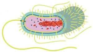
Prokaryotic cells lack a true nucleus and membrane-bound organelles.
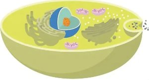
Eukaryotic cells possess a nucleus and various membrane-bound organelles.
Shapes and Arrangements of Prokaryotic Cells
Bacterial Morphology
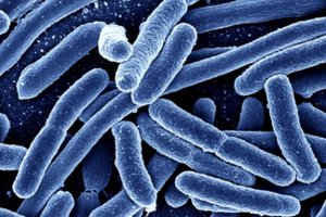
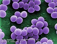
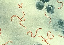
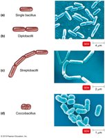
Bacteria exhibit a variety of shapes and arrangements, determined by their genetics. The three primary shapes are:
Bacillus (bacilli): Rod-shaped
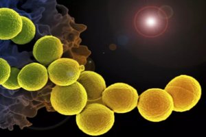
Coccus (cocci): Spherical
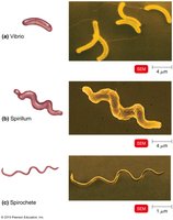
Spiral: Corkscrew-shaped (includes vibrio, spirillum, and spirochete)
Arrangements
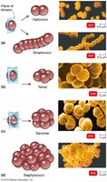
Staphylo-: Grape-like clusters (e.g., Staphylococcus)
Strepto-: Chains (e.g., Streptococcus)
Other Arrangements
Bacteria can also be found in pairs (diplococci, diplobacilli), tetrads, sarcinae (cubical packets), and palisades (side-by-side arrangement).
Prokaryotic Cell Structure
Major Components
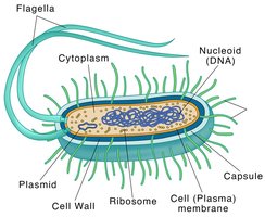
Cell wall: Provides shape and protection
Plasma membrane: Regulates entry and exit of substances
Cytoplasm: Gel-like substance inside the cell
Nucleoid: Region containing DNA
Ribosomes: Sites of protein synthesis
Plasmids: Small, circular DNA molecules
Capsule: Outer protective layer (if present)
Flagella, fimbriae, pili: Appendages for movement and attachment
Glycocalyxb
Structure and Types
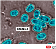
The glycocalyx is a viscous, gelatinous layer external to the cell wall, composed of polysaccharides and/or polypeptides. It exists in two forms:
Capsule: Neatly organized and firmly attached
Slime layer: Unorganized and loosely attached
Functions of Glycocalyx
Adherence: Helps bacteria attach to surfaces and each other (biofilm formation)
Virulence: Capsules prevent phagocytosis by host cells
Protection: Shields against dehydration and toxins
Nutrition: May serve as a nutrient reserve
Flagella and Motility
Flagella Structure
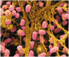
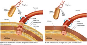
Flagella are long, whip-like appendages used for movement. They differ structurally between Gram-positive and Gram-negative bacteria.
Flagella Arrangements
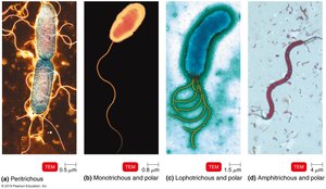
Monotrichous: Single flagellum at one end
Lophotrichous: Tuft of flagella at one end
Amphitrichous: Flagella at both ends
Peritrichous: Flagella distributed over the entire cell
Flagella Movement
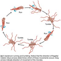
Bacteria move by rotating their flagella, a process called taxis. Movement can be toward (positive taxis) or away from (negative taxis) a stimulus, such as chemicals (chemotaxis).
Other Appendages
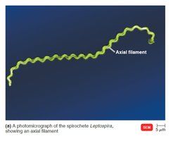
Axial filaments (endoflagella): Found in spirochetes, these structures wrap around the cell and enable corkscrew movement.
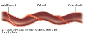
Fimbriae and Pili
Fimbriae: Short, hairlike structures for attachment
Pili: Longer, involved in motility and DNA transfer (conjugation)
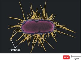
Bacterial Cell Wall
Structure and Function
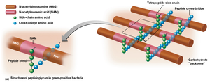
The bacterial cell wall maintains cell shape, prevents osmotic lysis, and anchors flagella. It is primarily composed of peptidoglycan, a polymer of sugars and amino acids.
Contains both D- and L-amino acids
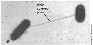
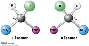
Gram-Positive vs. Gram-Negative Cell Walls
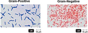
Bacteria are classified based on their cell wall structure, as revealed by the Gram stain:
Gram-positive: Thick peptidoglycan layer, teichoic acids
Gram-negative: Thin peptidoglycan layer, outer membrane with lipopolysaccharides, porins, and a periplasmic space
Plasma Membrane
Structure and Function
The plasma membrane is a phospholipid bilayer with embedded proteins, separating the cell wall from the cytoplasm. It is selectively permeable and involved in nutrient breakdown and energy generation.
Phosphate heads are hydrophilic; fatty acid tails are hydrophobic
Movement of Substances Across Membranes
Passive processes: Simple diffusion, facilitated diffusion, osmosis (no ATP required)
Active processes: Active transport, group translocation (require ATP)
Internal Structures of Prokaryotic Cells
Inclusions
Metachromatic granules: Inorganic phosphate storage
Polysaccharide and lipid inclusions: Energy reserves
Sulfur granules: Energy source for sulfur bacteria
Carboxysomes: Contain enzymes for photosynthesis
Gas vacuoles: Buoyancy in aquatic bacteria
Magnetosomes: Contain iron oxide, act as magnets
Endospores
Endospores are highly durable, dehydrated cells formed by some Gram-positive bacteria. They are resistant to heat, dehydration, chemicals, and radiation, and can survive for millions of years.
Eukaryotic Cell Structure
Major Components
Nucleus: Contains genetic material
Endoplasmic reticulum (ER): Rough ER (protein synthesis), Smooth ER (lipid synthesis)
Golgi complex: Protein modification and sorting
Lysosomes: Digestive enzymes
Vacuoles: Storage
Mitochondria: ATP production
Chloroplasts (plants/algae): Photosynthesis
Peroxisomes: Oxidation of fatty acids and detoxification
Centrosomes: Cell division
Flagella and Cilia
Eukaryotic flagella and cilia are projections for motility or moving substances along the cell surface. They have a 9+2 arrangement of microtubules and move in a wave-like manner.
Cell Wall and Glycocalyx
Cell wall: Present in plants, algae, fungi; made of cellulose, chitin, glucan, or mannan
Glycocalyx: Sticky carbohydrate layer for protection and attachment
Endosymbiotic Theory
The endosymbiotic theory proposes that eukaryotic cells evolved when larger prokaryotic cells engulfed smaller ones, which became organelles such as mitochondria and chloroplasts.
