 Back
BackFunctional Anatomy of Prokaryotic Cells: Structure and Function
Study Guide - Smart Notes
 Tailored notes based on your materials, expanded with key definitions, examples, and context.
Functional Anatomy of Prokaryotic Cells
Overview of Prokaryotic and Eukaryotic Cell Structure
Prokaryotic cells, such as bacteria and archaea, possess distinct structural features that differentiate them from eukaryotic cells. Understanding these differences is fundamental to microbiology.
Prokaryotes: One circular chromosome (not in a membrane), histone-like proteins, no organelles, peptidoglycan cell walls (bacteria), pseudomurein cell walls (archaea), 70S ribosomes, binary fission.
Eukaryotes: Paired chromosomes (in nuclear membrane), histones, organelles, polysaccharide cell walls (plants/fungi), no cell walls (animal cells), 80S ribosomes, mitosis.
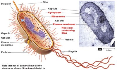
Bacterial Cell Structure
Bacterial cells contain several key structures, each with specific functions essential for survival and adaptation.
Capsule: Protective, often carbohydrate-rich layer outside the cell wall.
Cell Wall: Provides structural support and protection.
Plasma Membrane: Selectively permeable barrier, site of metabolic activity.
Nucleoid: Region containing the bacterial chromosome (DNA).
Plasmid: Small, circular DNA molecules carrying non-essential genes.
Ribosomes: Sites of protein synthesis (70S type).
Flagella, Pili, Fimbriae: External structures for motility and attachment.
Inclusions: Storage granules for nutrients.
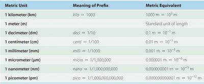
Bacterial Cell Size and Metric Units
Bacterial cells are typically measured in micrometers (µm), and understanding metric units is crucial for interpreting microscopic observations.
Average bacterial size: 0.2–1.0 µm × 2–8 µm.
Cellular organisms smaller than 0.15 µm are unlikely.
Metric Unit | Meaning of Prefix | Metric Equivalent |
|---|---|---|
Kilometer (km) | kilo = 1000 | 1000 m = m |
Meter (m) | Standard unit | 1 m |
Decimeter (dm) | deci = 1/10 | 0.1 m = m |
Centimeter (cm) | centi = 1/100 | 0.01 m = m |
Millimeter (mm) | milli = 1/1000 | 0.001 m = m |
Micrometer (µm) | micro = 1/1,000,000 | 0.000001 m = m |
Nanometer (nm) | nano = 1/1,000,000,000 | 0.000000001 m = m |
Picometer (pm) | pico = 1/1,000,000,000,000 | 0.000000000001 m = m |
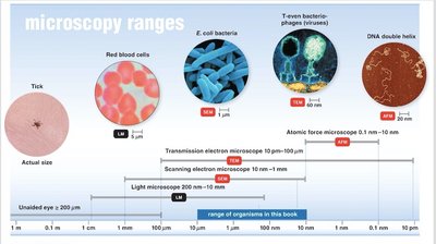
Microscopy Ranges
Different types of microscopes are used to observe microorganisms and cellular structures, each with specific resolution capabilities.
Light microscope: 200 nm–10 mm (cells, bacteria).
Transmission electron microscope (TEM): 10 pm–100 µm (viruses, organelles).
Scanning electron microscope (SEM): 1 nm–1 mm (surface structures).
Atomic force microscope (AFM): 0.1 nm–10 nm (molecular details).
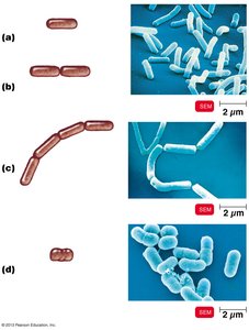
Bacterial Cell Shape and Arrangement
Common Shapes of Bacteria
Bacteria exhibit a variety of shapes, which are important for identification and classification.
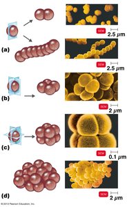
Bacillus: Rod-shaped (e.g., E. coli).
Coccus: Spherical-shaped (e.g., Streptococcus pneumoniae).
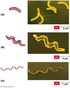
Spiral: Includes vibrio, spirillum, and spirochete (e.g., V. cholerae, Treponema pallidum).
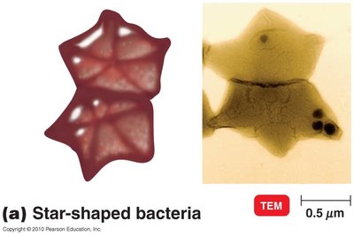
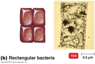
Star-shaped and Rectangular: Unusual forms.
Bacterial Plasma Membrane
Structure of the Plasma Membrane
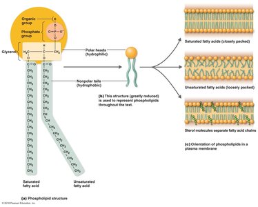
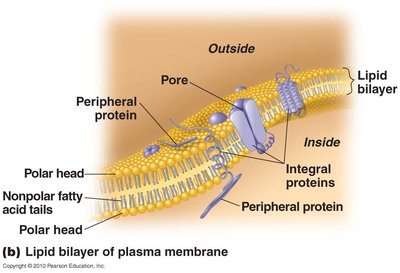
The plasma membrane is a phospholipid bilayer with embedded proteins, providing a selectively permeable barrier and facilitating cellular processes.
Phospholipids: Amphipathic molecules with hydrophilic heads and hydrophobic tails.
Fluid Mosaic Model: Proteins and lipids move freely; membrane is flexible and self-sealing.
Proteins: Peripheral, integral, and transmembrane proteins serve structural and functional roles.
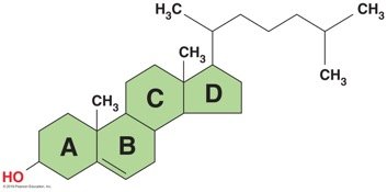
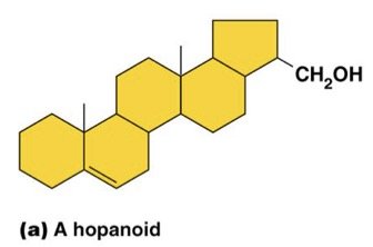
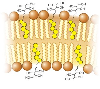
Membrane Lipids: Hopanoids and Sterols
Bacterial membranes contain hopanoids, which stabilize the membrane, while eukaryotic membranes contain sterols such as cholesterol.
Hopanoids: Unique to bacteria, functionally similar to cholesterol.
Cholesterol: Found in animal and plant membranes, decreases permeability.

Functions of the Plasma Membrane
The plasma membrane is essential for maintaining cellular integrity and mediating transport and energy production.
Encloses cytoplasm: Damage leads to leakage of cell contents.
Selectively permeable: Allows passage of some molecules, restricts others.
ATP production: Contains enzymes for electron transport chain.
Photosynthetic pigments: Present in some bacteria on chromatophores.
Anchors external structures: Flagella, pili, fimbriae.
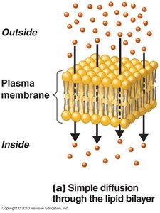
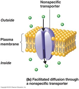
Movement of Materials Across the Plasma Membrane
Transport across the membrane occurs via passive and active processes, each with distinct mechanisms and energy requirements.
Passive Processes: No energy required; substances move from high to low concentration.
Active Processes: Energy required; substances move from low to high concentration.
Passive Processes
Simple Diffusion: Movement of solutes until equilibrium is reached.
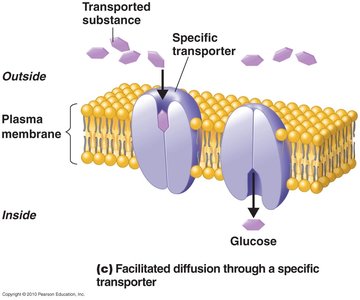
Facilitated Diffusion: Solutes combine with transporter proteins; transports ions and larger molecules.
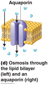
Osmosis: Movement of water across a selectively permeable membrane.

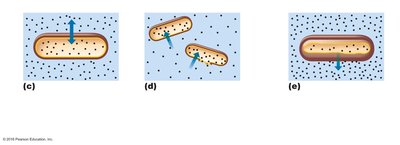
Tonicity and Osmotic Effects
Isotonic: Equal solute concentrations; no net water movement.
Hypotonic: Lower solute outside; water enters cell, may cause lysis.
Hypertonic: Higher solute outside; water leaves cell, causes plasmolysis.
Active Processes
Active Transport: Requires transporter protein and ATP; moves substances against gradient.
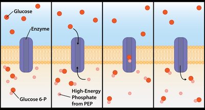
Group Translocation: Requires transporter protein and phosphoenolpyruvic acid (PEP); substance is chemically altered during transport.
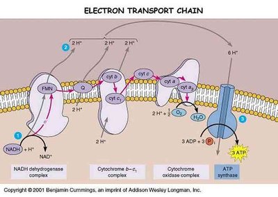
Energy Production in the Plasma Membrane
Bacterial plasma membranes contain enzymes for ATP production, primarily through the electron transport chain.
Electron Transport Chain: Series of protein complexes that transfer electrons and generate ATP.
 ----------------------------------------
