 Back
BackFunctional Anatomy of Prokaryotic Cells: Structure and Function
Study Guide - Smart Notes
 Tailored notes based on your materials, expanded with key definitions, examples, and context.
Functional Anatomy of Prokaryotic Cells
Overview of Prokaryotes and Eukaryotes
Prokaryotes and eukaryotes differ fundamentally in their cellular structure and organization. Understanding these differences is essential for microbiology students, as it forms the basis for classification and functional analysis of microorganisms.
Prokaryotes: Possess a single circular chromosome not enclosed in a membrane, lack organelles, have histone-like proteins, and typically have peptidoglycan (bacteria) or pseudomurein (archaea) cell walls. Ribosomes are 70S, and cell division occurs by binary fission.
Eukaryotes: Have paired chromosomes within a nuclear membrane, contain histones, organelles, and polysaccharide cell walls (plants and fungi). Animal cells lack cell walls. Ribosomes are 80S, and cell division occurs by mitosis.
Example: Escherichia coli is a typical prokaryote, while yeast (Saccharomyces cerevisiae) is a eukaryote.
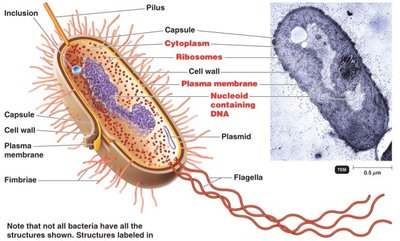
Bacterial Cell Structure
Bacterial cells exhibit a variety of structural components, each with specific functions. Not all bacteria possess every structure shown.
Capsule: Protective layer outside the cell wall, often involved in pathogenicity.
Cell wall: Provides structural support and shape; composed of peptidoglycan in bacteria.
Plasma membrane: Selectively permeable barrier, site of metabolic activities.
Nucleoid: Region containing the bacterial chromosome (DNA).
Ribosomes: Sites of protein synthesis (70S type).
Plasmid: Small, circular DNA molecules with additional genetic information.
Flagella, pili, fimbriae: External structures for motility and attachment.
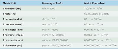
Metric Units and Microscopy Ranges
Microbiology requires understanding of metric units and the scale of microorganisms. The metric system is used to measure cell size, and microscopy techniques are essential for observation.
Metric units: Microorganisms are typically measured in micrometers (µm) and nanometers (nm).
Microscopy: Light microscopes can resolve cells down to 200 nm, while electron microscopes can visualize structures at the nanometer scale.
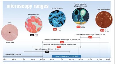
Bacterial Cell Size
Bacterial cells are generally small, with most ranging from 0.2–1.0 µm in width and 2–8 µm in length. Organisms smaller than 0.15 µm are unlikely to be cellular.
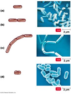
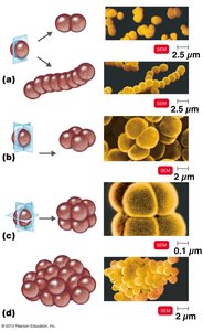
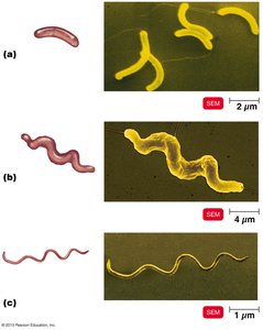
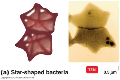
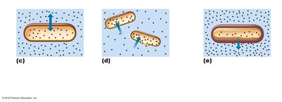
Bacterial Cell Shape
Bacteria display a variety of shapes, which are important for identification and classification. Most bacteria are monomorphic (single shape), but some are pleomorphic (variable shapes).
Bacillus: Rod-shaped; can be single, paired (diplo), chains (streptobacilli), or coccobacilli.
Coccus: Spherical; arrangements include diplo, strepto (chains), tetrads, sarcina (cubical), and staphylo (clusters).
Spiral: Includes vibrio (curved), spirillum (rigid spiral), and spirochete (flexible spiral).
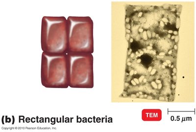
Unusual shapes: Star-shaped and rectangular bacteria exist but are rare.
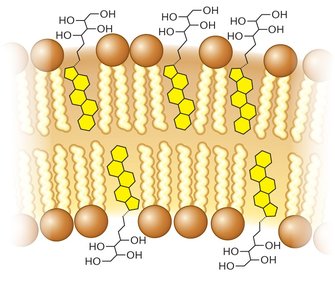
Plasma Membrane Structure
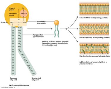
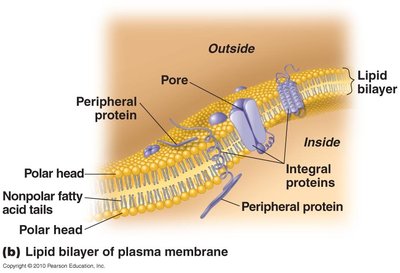
The plasma membrane is a critical structure in bacterial cells, composed primarily of phospholipids and proteins. It is amphipathic, with hydrophilic heads and hydrophobic tails, forming a bilayer.
Fluid mosaic model: Lipids and proteins move freely, allowing flexibility and self-sealing properties.
Proteins: Peripheral proteins are on the surface; integral and transmembrane proteins span the membrane.
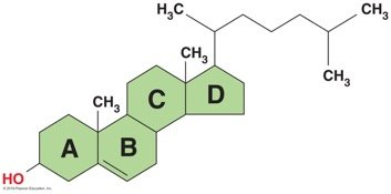
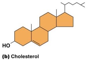
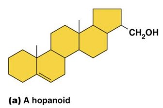
Hopanoids: Sterol-like molecules in bacterial membranes, providing stability.
Functions of the Plasma Membrane
The plasma membrane serves several essential functions in bacterial cells:
Encloses cytoplasm: Maintains cell integrity; damage leads to leakage.
Selective permeability: Allows passage of certain molecules while restricting others.
ATP production: Contains enzymes for electron transport and ATP synthesis.
Photosynthetic pigments: Present in some bacteria on membrane foldings (chromatophores).
Anchoring structures: Flagella, pili, and fimbriae are anchored in the membrane.
Movement of Materials Across the Plasma Membrane
Transport across the plasma membrane occurs via passive and active processes:
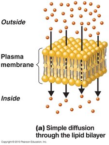
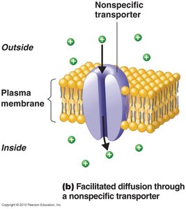
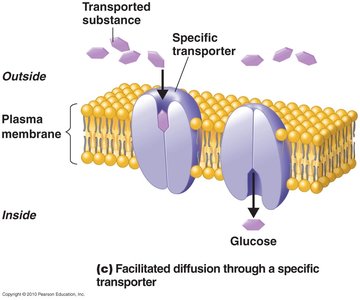
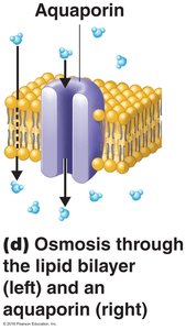
Passive processes: Do not require energy; include simple diffusion, facilitated diffusion, and osmosis.
Active processes: Require energy; include active transport and group translocation.
Passive Processes
Simple diffusion: Movement of solutes from high to low concentration until equilibrium is reached.
Facilitated diffusion: Solutes combine with transporter proteins to cross the membrane.
Osmosis: Movement of water across a selectively permeable membrane, either through the lipid bilayer or via aquaporins.
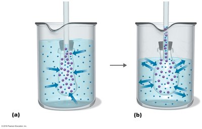
Tonicity and Osmosis
Isotonic solution: Equal solute concentration inside and outside; no net water movement.
Hypotonic solution: Lower solute concentration outside; water enters cell, may cause lysis.
Hypertonic solution: Higher solute concentration outside; water leaves cell, causing plasmolysis.
Active Processes
Active transport: Uses transporter proteins and ATP to move substances against concentration gradients.
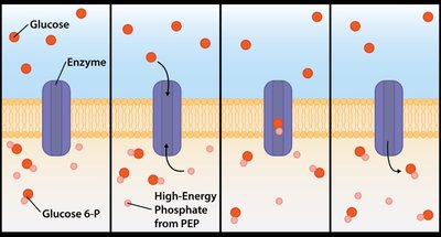
Group translocation: Substance is chemically modified during transport, often using phosphoenolpyruvic acid (PEP).
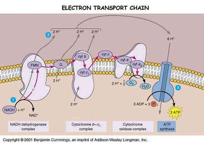
Energy Production in Bacteria
Bacterial plasma membranes contain enzymes for ATP production, primarily through the electron transport chain.
Photosynthesis in Bacteria
Some bacteria possess photosynthetic pigments on membrane foldings called chromatophores or thylakoids, enabling light-driven energy production.
Bacterial Cell Wall Structure
The bacterial cell wall is a defining feature, providing structural support and protection. It is composed of peptidoglycan in bacteria, with differences between Gram-positive and Gram-negative types.
Gram-positive: Thick peptidoglycan layer (20–30 layers).
Gram-negative: Thin peptidoglycan layer (1–2 layers) and an outer membrane.
Gram staining is used to differentiate these types based on cell wall structure.
