 Back
BackFunctional Anatomy of Prokaryotic Cells: Structure, Morphology, and External Features
Study Guide - Smart Notes
 Tailored notes based on your materials, expanded with key definitions, examples, and context.
Prokaryotic and Eukaryotic Cells
Basic Components of All Cells
All living cells share several fundamental structures that are essential for life. These include the plasma (cell) membrane, chromosomes, ribosomes, and cytosol.
Plasma (Cell) Membrane: A phospholipid bilayer that separates the living cell from its environment.
Chromosomes: DNA molecules carrying genetic information.
Ribosomes: Sites of protein synthesis.
Cytosol: Semi-fluid substance inside the cell membrane, primarily water.
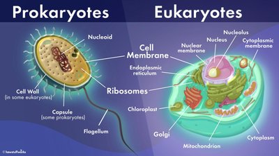
Differences Between Prokaryotic and Eukaryotic Cells
Prokaryotes and eukaryotes differ in several key structural and functional aspects:
Prokaryotes: Lack a membrane-bound nucleus; DNA is circular and not enclosed by a membrane. No membrane-bound organelles. Cell walls (if present) contain peptidoglycan (bacteria) or pseudomurein (archaea).
Eukaryotes: Have a true nucleus surrounded by a nuclear membrane. DNA is linear and organized with histones. Contain membrane-bound organelles. Cell walls (if present) are made of polysaccharides (e.g., chitin in fungi, cellulose in plants).
Cell replication: Prokaryotes divide by binary fission; eukaryotes use mitosis and a mitotic spindle.
Prokaryotic Cell Morphology and Arrangements
Shapes of Prokaryotic Cells
Bacteria exhibit a variety of shapes, which are important for classification and identification.
Coccus (plural: cocci): Spherical-shaped bacteria.
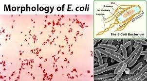
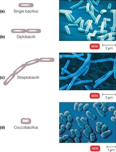
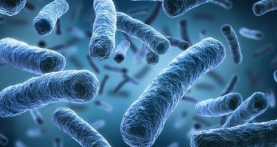
Bacillus (plural: bacilli): Rod-shaped bacteria.
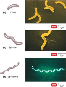
Spiral: Includes vibrio (curved rods), spirillum (rigid helix), and spirochete (flexible helix).
Pleomorphic: Bacteria that can have variable shapes due to genetic or environmental factors.
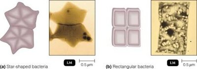
Unusual shapes: Some bacteria are star-shaped (genus Stella) or rectangular (genus Haloarcula).
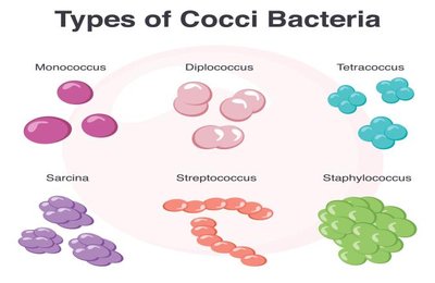
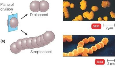
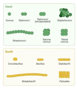
Bacterial Arrangements
Bacteria can be arranged in specific patterns based on their division and attachment:
Pairs: Diplococci, diplobacilli
Chains: Streptococci, streptobacilli
Clusters: Staphylococci (not found in bacilli due to their division pattern)
Tetrads, sarcinae: Groups of four or eight cocci, respectively
Structure of Prokaryotic Cells
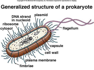
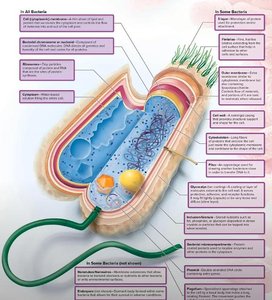
Generalized Structure
Prokaryotic cells have several essential and accessory structures that contribute to their survival and function.
Essential structures (in all bacteria): Cell membrane, nucleoid (bacterial chromosome), ribosomes, cytoplasm
Accessory structures (in some bacteria): S layer, fimbriae, outer membrane, cell wall, cytoskeleton, pilus, glycocalyx, inclusions/granules, microcompartments, plasmids, flagellum
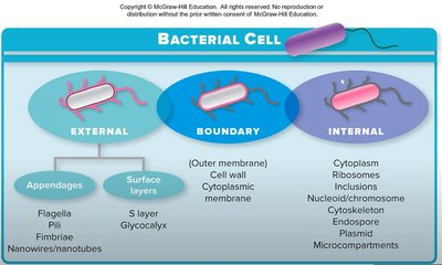
External Structures: Glycocalyx
Types and Functions
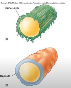
The glycocalyx is a gelatinous external layer composed of polysaccharide or polypeptide. It exists in two forms:
Slime layer: Loosely organized and attached.
Capsule: Highly organized and tightly attached.
Functions of the glycocalyx include:
Prevention of dehydration and nutrient loss
Adherence to surfaces and biofilm formation
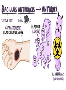
Protection from phagocytosis (e.g., Bacillus anthracis capsule made of D-glutamic acid)
Virulence factor: enhances the ability to cause disease (e.g., Streptococcus pneumoniae)
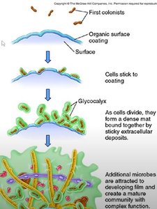
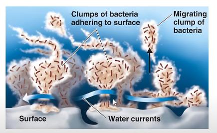
Biofilm Formation
Bacteria with a glycocalyx can form biofilms—complex, surface-attached microbial communities. Biofilms provide protection from antibiotics, immune responses, and environmental stresses.
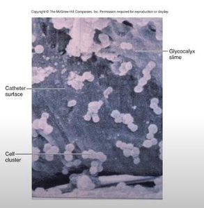
Biofilms are common on teeth, mucus membranes, heart valves, catheters, and implants.
Biofilm bacteria communicate via quorum sensing (chemical signaling).
Biofilms facilitate nutrient sharing and genetic exchange (e.g., conjugation).
Motility and Attachment Structures
Flagella
Flagella are long, whip-like appendages used for bacterial motility. They consist of three main parts:
Filament: Composed of flagellin protein, forms a helical structure.
Hook: Connects the filament to the basal body and allows rotation.
Basal body: Anchors the flagellum to the cell wall and membrane; structure differs in Gram-positive and Gram-negative bacteria.
Flagellar arrangements include monotrichous (single), lophotrichous (tuft at one end), amphitrichous (tufts at both ends), and peritrichous (all over the cell). Movement is achieved by rotation: counterclockwise for runs (directed movement), clockwise for tumbles (change in direction).
Flagella also serve as H antigens for bacterial identification (e.g., E. coli O157:H7).
Axial Filaments
Axial filaments (endoflagella) are found in spirochetes and allow corkscrew movement, aiding in tissue penetration (e.g., Treponema pallidum causes syphilis).
Fimbriae and Pili
Fimbriae: Short, hair-like structures for attachment to surfaces and host tissues; not involved in motility.
Pili: Longer, rigid structures involved in conjugation (DNA transfer), attachment, and sometimes motility.
Internal Structures of Prokaryotes
Cytoplasm
The cytoplasm is a gel-like matrix composed of water, proteins, carbohydrates, lipids, and ions. It serves as a pool for building blocks and energy sources.
Nucleoid and Plasmids
Nucleoid: Region containing the bacterial chromosome (single, circular, double-stranded DNA).
Plasmids: Small, circular DNA molecules carrying non-essential genes (e.g., antibiotic resistance).
Ribosomes
Prokaryotic ribosomes are 70S (composed of 50S and 30S subunits) and are the sites of protein synthesis.
Inclusions
Inclusions are storage granules for nutrients and metabolic by-products. Gas vacuoles provide buoyancy for aquatic bacteria (e.g., cyanobacteria) to perform photosynthesis.
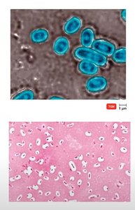
Endospores
Endospores are highly resistant, dormant structures formed by some Gram-positive bacteria (e.g., Bacillus, Clostridium) for survival under adverse conditions. They can withstand dehydration, heat, radiation, and chemicals. Endospore formation (sporulation) is not a reproductive process; germination returns the cell to a vegetative state when conditions improve.
Cell Wall Structure and Function
Peptidoglycan Structure
The bacterial cell wall is primarily composed of peptidoglycan, a polymer of alternating N-acetylglucosamine (NAG) and N-acetylmuramic acid (NAM) linked by β-1,4-glycosidic bonds and cross-linked by peptides.
Gram-positive cell walls: Thick peptidoglycan layer, teichoic acids (for cation regulation and antigenicity), no outer membrane.
Gram-negative cell walls: Thin peptidoglycan layer, outer membrane containing lipopolysaccharides (LPS), lipoproteins, and phospholipids. The periplasmic space lies between the outer and plasma membranes.
LPS contains O-polysaccharide (antigenic), core polysaccharide (stability), and Lipid A (endotoxin).
Gram Staining
Gram staining differentiates bacteria based on cell wall structure:
Gram-positive: Retain crystal violet stain (purple) due to thick peptidoglycan.
Gram-negative: Lose crystal violet, retain safranin (pink) due to thin peptidoglycan and outer membrane.
Atypical Cell Walls
Mycobacterium: Waxy, mycolic acid-rich cell wall; acid-fast, highly resistant to chemicals and dehydration.
Mycoplasma: Lack cell wall; pleomorphic, sensitive to osmotic lysis, contain sterols in the membrane for stability.
Cell Wall Damage and Antibiotics
Lysozyme: Enzyme that digests peptidoglycan, found in body fluids.
Penicillin: Inhibits peptide cross-bridge formation in peptidoglycan, more effective against Gram-positive bacteria.
Gram-negative bacteria are more resistant to antibiotics due to their outer membrane and porins.
Summary Table: Key Differences Between Gram-Positive and Gram-Negative Bacteria
Feature | Gram-Positive | Gram-Negative |
|---|---|---|
Peptidoglycan Layer | Thick | Thin |
Teichoic Acids | Present | Absent |
Outer Membrane | Absent | Present (LPS, porins) |
Sensitivity to Penicillin | High | Low |
Lipid A (Endotoxin) | Absent | Present |
Gram Stain Color | Purple | Pink |
Additional info: The notes above integrate and expand upon the provided lecture content, ensuring a comprehensive, exam-ready summary of prokaryotic cell structure and function, with relevant images included only where they directly reinforce the text.
