 Back
Immune System Disorders and Hypersensitivity Reactions
Study Guide - Smart Notes
 Tailored notes based on your materials, expanded with key definitions, examples, and context.
Immune System Disorders
Overview of Immune Deficiencies
The immune system is essential for defending the body against pathogens and abnormal cells. Immune deficiencies occur when this system fails to function properly, leading to increased susceptibility to infections and, in some cases, cancer. Immune deficiencies are classified as either primary (genetic) or secondary (acquired).
Primary immunodeficiency: Caused by genetic defects, often present from birth, and may affect B cells, T cells, phagocytes, or complement proteins.
Secondary immunodeficiency: Acquired later in life due to factors such as aging, infections (e.g., HIV), medical treatments, or chronic diseases.
Primary Immunodeficiencies
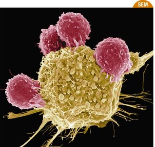
Primary immunodeficiencies are rare, lifelong disorders resulting from inborn errors that impair one or more components of the immune system. Over 300 types have been identified, with varying severity and treatment options.
B cell/antibody deficiencies: e.g., Selective IgA deficiency, X-linked agammaglobulinemia
T cell deficiencies: e.g., DiGeorge syndrome, congenital thymic dysplasia
Combined immunodeficiencies: e.g., Severe combined immunodeficiency (SCID), Wiskott-Aldrich syndrome
Phagocyte defects: e.g., Chronic granulomatous disease
Complement deficiencies: e.g., C2, C4, C9 deficiencies
Treatment options include bone marrow transplants, antibody administration, cytokine therapies, and experimental approaches such as gene therapy.
Secondary Immunodeficiencies
Secondary immunodeficiencies are more common and result from external factors that weaken the immune system after birth. Causes include:
Aging
Infectious agents (e.g., HIV)
Medical interventions (e.g., immunosuppressive drugs)
Chronic diseases (e.g., diabetes, malnutrition, alcoholism)
Some drugs, such as corticosteroids and immunosuppressants, can induce immunodeficiency, which may be reversible if the drug is discontinued.
Immunodeficiency and Cancer
The immune system also plays a role in cancer surveillance. Immunodeficient individuals, such as those with HIV/AIDS or organ transplant recipients, have a higher risk of developing certain cancers.
Autoimmune Disorders
Definition and Mechanisms
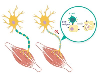
Autoimmunity occurs when the immune system mistakenly attacks the body's own healthy tissues, leading to chronic conditions known as autoimmune disorders. The loss of self-tolerance is central to the development of these diseases.
Genetic predisposition and environmental triggers (e.g., infections) contribute to autoimmunity.
Autoimmune diseases can be systemic (affecting multiple organs) or localized (targeting specific tissues).
Examples of Autoimmune Diseases
Type 1 diabetes mellitus: Immune attack on pancreatic beta cells
Multiple sclerosis: Immune-mediated damage to myelin in the nervous system
Rheumatoid arthritis: Immune attack on joint linings
Systemic lupus erythematosus (SLE): Systemic attack on DNA, histones, and other nuclear components
Guillain-Barré syndrome: Immune attack on peripheral nerves
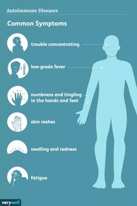
Diagnosis involves detecting self-reactive immune cells or autoantibodies, along with clinical symptoms such as joint pain, fatigue, rash, and organ dysfunction.
Hypersensitivity Reactions
Definition and Classification
Hypersensitivities are inappropriate or exaggerated immune responses to antigens, which can be either localized or systemic. The Gell and Coombs classification system divides hypersensitivities into four types:
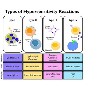
Type | Mechanism | Onset | Examples |
|---|---|---|---|
I (Immediate) | IgE-mediated, mast cell degranulation | Within 1 hour | Anaphylaxis, allergies |
II (Cytotoxic) | IgG/IgM-mediated, cell lysis | Hours to days | Hemolytic anemia, transfusion reactions |
III (Immune Complex) | Immune complex deposition, complement activation | 1-3 weeks | Serum sickness, SLE |
IV (Delayed) | T cell-mediated, cytokine release | Days to weeks | Contact dermatitis, MS |
Type I Hypersensitivity (Immediate, IgE-Mediated)
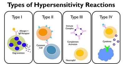
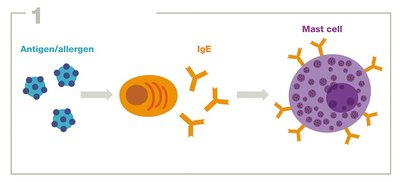
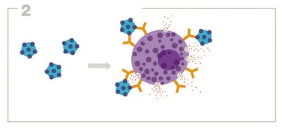
Type I hypersensitivity involves IgE antibodies binding to allergens, leading to mast cell or basophil degranulation and release of inflammatory mediators. This type includes most allergies and anaphylaxis.
Sensitization: Initial exposure to allergen triggers IgE production, which binds to mast cells.
Subsequent exposure: Allergen cross-links IgE on mast cells, causing degranulation and release of histamine.
Symptoms: Range from mild (hives, rhinitis) to severe (anaphylactic shock).
Diagnosis and Management
Blood tests: Measure allergen-specific IgE levels.
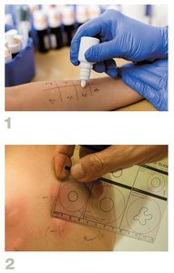
Skin prick tests: Identify specific allergens by observing wheal and flare reactions.
Desensitization therapy: Gradual exposure to increasing amounts of allergen to reduce sensitivity (effective for some non-food allergies).
Type II Hypersensitivity (Cytotoxic, IgG/IgM-Mediated)
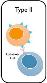
Type II hypersensitivity involves IgG or IgM antibodies binding to antigens on cell surfaces, leading to cell destruction via complement activation or recruitment of cytotoxic cells.
Examples: Hemolytic disease of the newborn (Rh incompatibility), blood transfusion reactions, some autoimmune diseases (e.g., Goodpasture syndrome).
Mechanisms: Complement-dependent cytolysis, antibody-dependent cell-mediated cytotoxicity (ADCC), and opsonization for phagocytosis.
Blood Groups and Transfusion Reactions
Blood type compatibility is determined by the presence of A, B, and Rh antigens on red blood cells. Incompatible transfusions can cause rapid and severe hemolytic reactions.
Type III Hypersensitivity (Immune Complex-Mediated)
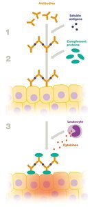
Type III hypersensitivity is caused by the formation of antigen-antibody complexes that deposit in tissues, activating complement and attracting leukocytes, leading to inflammation and tissue damage.
Examples: Systemic lupus erythematosus (SLE), rheumatoid arthritis, poststreptococcal glomerulonephritis, serum sickness.
Type IV Hypersensitivity (Delayed, T Cell-Mediated)
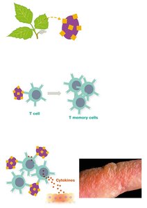
Type IV hypersensitivity is mediated by T cells rather than antibodies. The response is delayed, typically manifesting 12–72 hours after antigen exposure.
Examples: Contact dermatitis (e.g., poison ivy), tuberculin skin test, transplant rejection, autoimmune diseases such as type I diabetes and multiple sclerosis.
Mechanism: Sensitized T cells release cytokines, recruit phagocytes, and cause tissue inflammation and damage.
Summary Table: Hypersensitivity Types
Type | Immune Mediator | Onset | Examples |
|---|---|---|---|
I | IgE, mast cells | Minutes | Allergies, anaphylaxis |
II | IgG/IgM, complement | Hours to days | Transfusion reactions, HDN |
III | Immune complexes | 1–3 weeks | Lupus, serum sickness |
IV | T cells | Days | Contact dermatitis, MS |
Key Takeaways
Immune system disorders include immunodeficiencies and autoimmune diseases.
Hypersensitivity reactions are classified into four types based on immune mechanisms and clinical features.
Diagnosis and management of these disorders require understanding of underlying immunological principles.
