 Back
BackInnate and Adaptive Immunity: Mechanisms and Cellular Components
Study Guide - Smart Notes
 Tailored notes based on your materials, expanded with key definitions, examples, and context.
Overview of the Immune Response
Introduction to Immunity
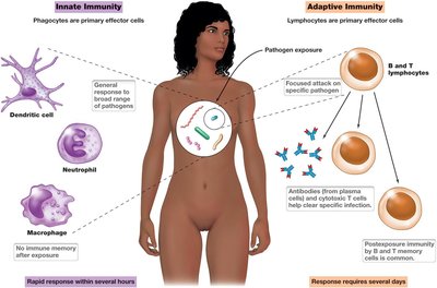
The immune system protects organisms from infection through a complex network of cells, tissues, and molecules. Immunity is the ability of an organism to resist infection, and it is divided into two main branches: innate (non-specific) and adaptive (specific) immunity.
Innate Immunity: Provides immediate, non-specific defense against pathogens.
Adaptive Immunity: Develops more slowly and targets specific pathogens with high precision.
Immune Response Cells and Hematopoiesis
Origin and Differentiation of Immune Cells
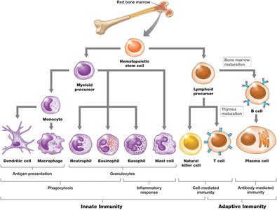
All immune cells originate from hematopoietic stem cells in the bone marrow. These stem cells differentiate into various leukocytes (white blood cells) that participate in both innate and adaptive immune responses.
Myeloid lineage: Produces phagocytes (e.g., neutrophils, macrophages, dendritic cells) and other cells involved in innate immunity.
Lymphoid lineage: Produces lymphocytes (B cells, T cells, and natural killer cells) involved in adaptive and innate immunity.
The Blood and Lymphatic Systems
Structure and Function
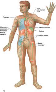
The blood and lymphatic systems are essential for the transport and communication of immune cells throughout the body. Lymphoid organs (e.g., thymus, spleen, lymph nodes) are sites of immune cell maturation and activation.
Primary lymphoid organs: Bone marrow and thymus (sites of immune cell development).
Secondary lymphoid organs: Lymph nodes, spleen, and mucosa-associated lymphoid tissue (MALT) (sites of immune response initiation).
Blood and Lymph Capillaries
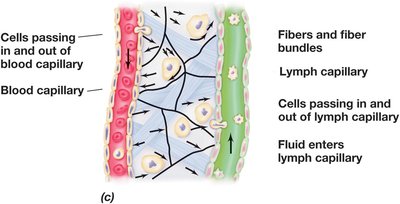
Immune cells circulate between blood and lymphatic vessels, allowing for efficient surveillance and response to pathogens.
Cells and fluid can pass in and out of both blood and lymph capillaries, facilitating immune cell migration to sites of infection.
Innate (Non-specific) Immunity
Key Cellular Components
Innate immunity is the first line of internal defense and involves several types of leukocytes:
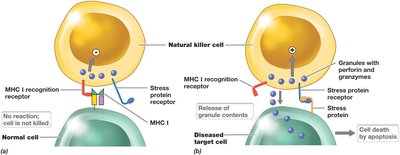
Natural Killer (NK) Cells: Recognize and kill virus-infected, cancerous, or stressed cells by releasing perforin and granzymes.
Phagocytes: Neutrophils, monocytes, macrophages, and dendritic cells engulf and destroy pathogens by phagocytosis.
Major Histocompatibility Complex (MHC) Proteins
MHC proteins are essential for distinguishing self from non-self and for antigen presentation:
MHC I: Present on all nucleated cells; recognized by NK cells and present antigens to cytotoxic T cells.
MHC II: Present on leukocytes (macrophages, dendritic cells, B cells); present antigens to T-helper cells.
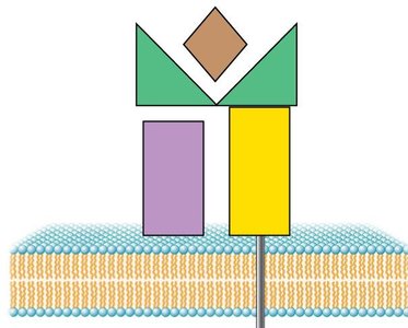
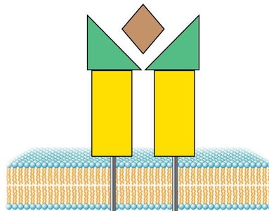

Recognition of Cells by Natural Killer Cells
NK cells distinguish healthy cells from infected or cancerous cells based on the presence or absence of MHC I molecules and stress proteins.
If MHC I is present, NK cells are deactivated.
If stress proteins are detected (often in infected or cancerous cells), NK cells induce apoptosis.
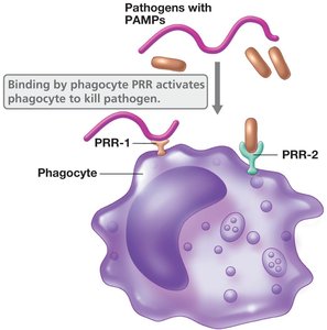
Phagocytosis and Pattern Recognition
Phagocytes recognize pathogens using pattern recognition receptors (PRRs) that bind pathogen-associated molecular patterns (PAMPs).
Binding of PRRs to PAMPs activates phagocytes to engulf and destroy pathogens.
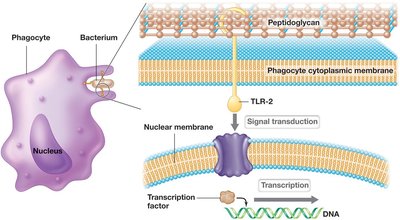
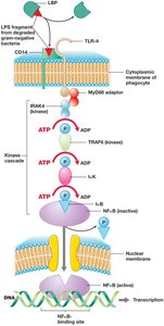
Toll-Like Receptors (TLRs) and Signal Transduction
TLRs are a type of PRR that recognize conserved microbial structures and initiate intracellular signaling cascades, leading to the activation of genes involved in inflammation and immune responses.
Signal transduction through TLRs results in the production of cytokines and other immune mediators.
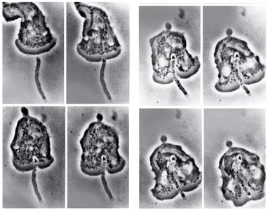
Phagocytosis in Action
Phagocytosis is the process by which phagocytes engulf and digest pathogens. Some pathogens have evolved mechanisms to evade phagocytosis, such as producing pigments, scavenging toxic oxygen, or forming capsules.
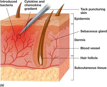
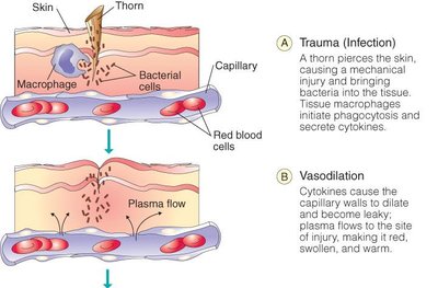
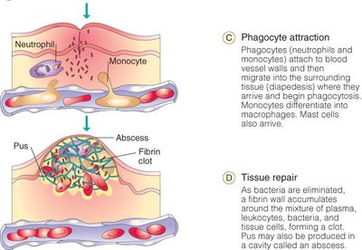
Inflammation
Mechanisms and Cellular Events
Inflammation is a localized, non-specific response to infection or injury, characterized by redness, heat, swelling, and pain. It is closely linked to phagocytosis and the recruitment of immune cells to the site of infection.
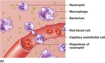
Neutrophils and macrophages are rapidly recruited to the site of injury by chemokines and cytokines.
These cells secrete inflammatory mediators (e.g., interleukins, TNF-α) that increase vascular permeability and attract additional immune cells.
Systemic Inflammatory Response and Septic Shock
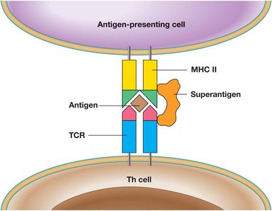
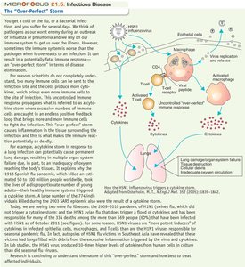
When inflammation becomes systemic, it can lead to life-threatening conditions such as septic shock, often triggered by the introduction of large numbers of bacteria or superantigens into sterile body spaces.
Superantigens can activate an abnormally large number of T cells, resulting in excessive cytokine production and systemic inflammation.
Adaptive (Specific) Immunity
Properties and Overview
Adaptive immunity is characterized by specificity, memory, and tolerance. It involves the recognition of specific antigens, the generation of memory cells for faster secondary responses, and the prevention of self-reactivity.
Specificity: Immune cells recognize and react with specific antigens.
Memory: Memory T and B cells enable rapid and robust responses upon re-exposure to the same antigen.
Tolerance: Immune cells do not react with self-antigens.
Types of Adaptive Immune Responses
Cell-mediated immunity: Involves T cells (T-cytotoxic and T-helper 1) and phagocytes.
Antibody-mediated (humoral) immunity: Involves B cells and T-helper 2 cells, leading to antibody production.
T-Cell Receptors and Selection
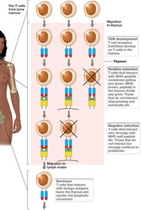
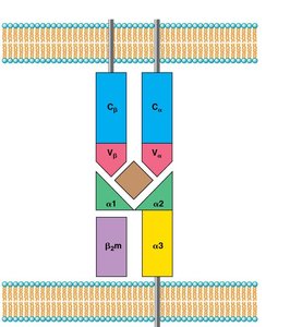
T-cell receptors (TCRs) are proteins on the surface of T cells that specifically bind antigen/MHC complexes. T cells undergo positive and negative selection in the thymus to ensure self-tolerance and functional competence.
Positive selection: T cells that can bind MHC proteins survive.
Negative selection: T cells that bind self-antigen/MHC complexes too tightly are eliminated.
Activation and Differentiation of T Cells
When a T-cell receptor binds to an antigen/MHC complex, the T cell becomes activated and divides to produce effector and memory cells.
Effector cells: Carry out immune functions and are short-lived.
Memory cells: Persist long-term and respond rapidly upon re-exposure to the antigen.
Cell-Mediated Immunity
Cell-mediated immunity is primarily carried out by T-cytotoxic (Tc) cells and T-helper 1 (Th1) cells.
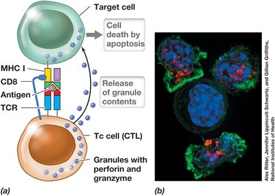
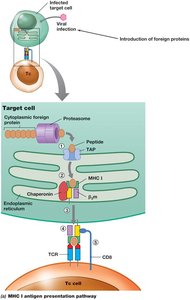
T-cytotoxic cells (CTLs): Kill cells displaying foreign antigens on MHC I proteins by releasing perforin and granzymes, inducing apoptosis.
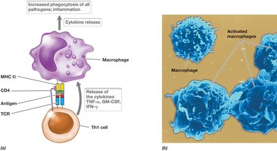
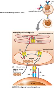
T-helper 1 cells (Th1): Activated by phagocytes presenting antigens on MHC II proteins; secrete cytokines to enhance phagocytosis and inflammation.
Antigen Presentation Pathways
Antigen presentation is essential for T cell activation and occurs via two main pathways:
MHC I pathway: Presents endogenous antigens to cytotoxic T cells.
MHC II pathway: Presents exogenous antigens to helper T cells.
Antibody-Mediated (Humoral) Immunity
Antibodies (Immunoglobulins)
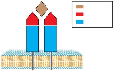
Antibodies are Y-shaped proteins produced by B cells that specifically bind antigens. There are five major classes of antibodies, each with distinct functions:
Class | Main Function |
|---|---|
IgG | Major antibody in serum; crosses placenta |
IgA | Predominant in secretions (saliva, breast milk, intestinal fluid) |
IgM | First antibody produced in response to infection |
IgD | Low concentration; functionally similar to IgM |
IgE | Low concentration; responsible for allergic reactions |
Immunoglobulin Gene Rearrangement
Somatic recombination in B cells generates a vast diversity of antibodies, allowing the immune system to recognize an almost unlimited variety of antigens.
Each B cell produces only one kind of antibody, but the total diversity is immense due to gene rearrangement.
Primary and Secondary Immune Responses
The primary immune response occurs upon first exposure to an antigen, leading to the production of antibodies and memory cells. The secondary response is faster and stronger due to the presence of memory cells.
Primary response: Involves initial antibody production (mainly IgM) and formation of memory cells.
Secondary response: Rapid production of high-affinity antibodies (mainly IgG) upon re-exposure to the antigen.
Destruction of Pathogens: The Complement System
Complement Activation Pathways
The complement system consists of serum proteins (C1–C9) that enhance the ability of antibodies and phagocytes to clear pathogens. There are two main activation pathways:
Classical pathway: Triggered by antigen-antibody complexes.
Alternative pathway: Triggered directly by pathogen surfaces.
Outcomes of Complement Activation
Opsonization: Coating of pathogens to enhance phagocytosis.
Cell lysis: Formation of the membrane attack complex (MAC) that creates pores in pathogen membranes.
Inflammation: Recruitment of immune cells and increased vascular permeability.
