 Back
BackInnate Immunity and Host Defenses – Microbiology Study Guidance
Study Guide - Smart Notes
 Tailored notes based on your materials, expanded with key definitions, examples, and context.
Q1. Which of the complement fragments is inflammatory?
Background
Topic: Complement System in Innate Immunity
This question tests your understanding of the complement cascade, specifically which fragments (C3a, C4a, C5a) are involved in inflammation.
Key Terms and Concepts:
Complement System: A group of proteins that enhance immune responses, including inflammation, opsonization, and cell lysis.
Inflammatory Fragments: Certain complement fragments act as chemotactic factors and increase vascular permeability, promoting inflammation.
Step-by-Step Guidance
Recall the three main outcomes of complement activation: opsonization, cell lysis, and inflammation.
Review the roles of C3a, C4a, and C5a in the complement cascade. Which ones are known as anaphylatoxins?
Think about which fragment is considered the most potent inflammatory mediator.
Consider the answer choices and eliminate any that do not have a strong role in inflammation.
Try solving on your own before revealing the answer!
Final Answer: d. all of the above
C3a, C4a, and C5a are all involved in inflammation, but C5a is the most potent anaphylatoxin. All three contribute to the inflammatory response.
Q2. The type of interferon present late in an infection is __________.
Background
Topic: Interferons in Antiviral Defense
This question is about the different types of interferons (alpha, beta, gamma, delta) and their roles during infection.
Key Terms and Concepts:
Interferons (IFNs): Cytokines that help regulate the immune response, especially against viruses.
Type I IFNs: (alpha and beta) produced early in viral infections.
Type II IFN: (gamma) produced later, mainly by T cells and NK cells.
Step-by-Step Guidance
Recall which interferons are produced first in response to viral infection (Type I: alpha and beta).
Think about which interferon is produced later and is associated with activation of macrophages and adaptive immunity.
Review the answer choices and match the interferon type to its timing in infection.
Try solving on your own before revealing the answer!
Final Answer: c. gamma interferon
Gamma interferon is produced later in infection and is important for activating macrophages and enhancing adaptive immunity.
Q3. True/False and Correction: The surface cells of the epidermis of the skin are __________.
Background
Topic: Physical Barriers in Innate Immunity
This question tests your knowledge of the structure and function of the skin as a barrier to infection.
Key Terms and Concepts:
Epidermis: The outermost layer of skin, composed of tightly packed cells.
Keratinization: Surface cells are often dead and filled with keratin, providing a tough barrier.
Step-by-Step Guidance
Recall the structure of the epidermis and whether the surface cells are alive or dead.
If the statement is false, correct it by stating the true condition of the surface cells.
Try solving on your own before revealing the answer!
Final Answer: False. The surface cells of the epidermis are dead.
The outermost cells are dead and filled with keratin, forming a protective barrier.
Q4. Match the following: Epidermis, Lysozyme, Goblet cells, Phagocytes, Sebum, T lymphocytes, Antimicrobial peptides
Background
Topic: Lines of Defense in Immunity
This question asks you to match immune components to the first, second, or third line of defense.
Key Terms and Concepts:
First Line of Defense: Physical and chemical barriers (e.g., skin, mucous membranes, lysozyme, sebum).
Second Line of Defense: Cellular defenses (e.g., phagocytes, antimicrobial peptides).
Third Line of Defense: Adaptive immunity (e.g., T lymphocytes).
Step-by-Step Guidance
Review the function of each component listed.
Assign each to the appropriate line of defense based on its role (barrier, innate cellular, or adaptive).
Double-check your matches by recalling examples of each defense line.
Try solving on your own before revealing the answer!
Final Answer:
Epidermis – First line
Lysozyme – First line
Goblet cells – First line
Phagocytes – Second line
Sebum – First line
T lymphocytes – Third line
Antimicrobial peptides – Second line
Each component is matched to the defense line based on its function in immunity.
Q5. Label the diagram of a phagocyte
Background
Topic: Phagocytosis and Immune Cells
This question asks you to identify structures involved in phagocytosis on a diagram of a phagocyte.
Key Terms and Concepts:
Phagocyte: A cell that engulfs and digests pathogens and debris.
Phagosome: Vesicle containing the ingested particle.
Lysosome: Organelle containing digestive enzymes.
Phagolysosome: Fusion of phagosome and lysosome for digestion.
Step-by-Step Guidance
Identify the cell membrane, ingested particle, phagosome, lysosome, and phagolysosome on the diagram.
Label each structure based on its appearance and location in the process of phagocytosis.
Use the diagram to reinforce your understanding of the steps of phagocytosis.
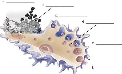
Try solving on your own before revealing the answer!
Final Answer:
a. Bacterium
b. Pseudopodia
c. Phagosome
d. Lysosome
e. Phagolysosome
f. Exocytosis of debris
These labels correspond to the main steps and structures involved in phagocytosis.
