 Back
BackInnate Immunity: Nonspecific Defenses of the Host – Study Notes
Study Guide - Smart Notes
 Tailored notes based on your materials, expanded with key definitions, examples, and context.
Innate Immunity: Nonspecific Defenses of the Host
Overview of Immunity
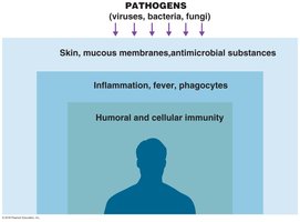
Immunity is the ability of an organism to resist infection or disease. The immune system is divided into innate (nonspecific) and adaptive (specific) immunity. Innate immunity provides the first and second lines of defense against pathogens, while adaptive immunity offers a targeted response.
Immunity: Ability to ward off disease.
Susceptibility: Lack of resistance to a disease.
Innate Immunity: Defenses against any pathogen; present from birth.
Adaptive Immunity: Immunity or resistance to a specific pathogen; involves memory.
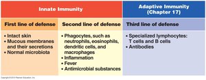
First Line of Defense: Skin and Mucous Membranes
Physical Barriers
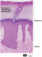
The first line of defense includes physical and chemical barriers that prevent pathogen entry. The skin and mucous membranes are primary physical barriers.
Skin: Consists of the dermis (inner connective tissue) and epidermis (outer epithelial cells with keratin). Shedding and dryness inhibit microbial growth.
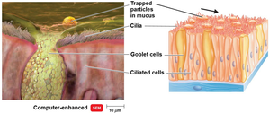
Mucous Membranes: Line the gastrointestinal, respiratory, and genitourinary tracts. Mucus traps microbes and prevents tract desiccation.
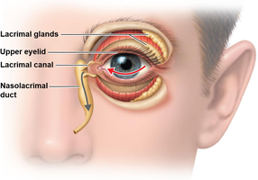
Lacrimal Apparatus: Produces tears that wash the eye, removing microbes.
Other Physical Factors
Ciliary Escalator: Moves mucus and trapped microbes away from the lungs.
Earwax: Prevents entry of microbes into the ear canal.
Urine Flow: Cleanses the urethra of microbes.
Vaginal Secretions: Remove microorganisms from the vaginal tract.
Peristalsis, Defecation, Vomiting, Diarrhea: Expel microbes from the body.
Chemical Barriers
Chemical factors enhance the effectiveness of physical barriers by creating hostile environments for pathogens.
Sebum: Forms a protective film and lowers skin pH (3–5).
Lysozyme: Enzyme in perspiration, tears, saliva, and urine that destroys bacterial cell walls.
Gastric Juice: Low pH (1.2–3.0) destroys most bacteria and toxins.
Vaginal Secretions: Low pH (3–5) inhibits microbes.
Role of Normal Microbiota
Normal microbiota protect the host by competing with pathogens (microbial antagonism), producing substances harmful to pathogens, and altering environmental conditions.
Commensalism: One organism benefits, the other is unharmed.
Probiotics: Live microbial cultures administered for beneficial effects.
Second Line of Defense: Cellular and Molecular Responses
Leukocytes (White Blood Cells)
Leukocytes are essential for innate immunity, identifying and eliminating pathogens through various mechanisms.
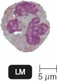
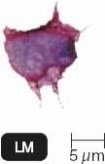
Neutrophils: Phagocytic; first responders to infection.
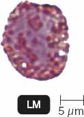
Basophils: Release histamine; involved in inflammation and allergic responses.
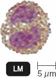
Eosinophils: Kill parasites and modulate allergic inflammatory responses.
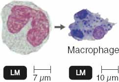
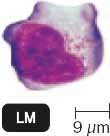
Monocytes: Differentiate into macrophages and dendritic cells in tissues; phagocytic.
Lymphocytes: Include natural killer (NK) cells, T cells, and B cells (adaptive immunity).
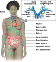
The Lymphatic System
The lymphatic system transports lymph, which contains lymphocytes and phagocytic cells, and filters pathogens through lymph nodes.
Lymph: Fluid containing immune cells.
Lymphoid Tissue: Includes lymph nodes, spleen, tonsils, and Peyer's patches.
Red Bone Marrow: Site of immune cell production.
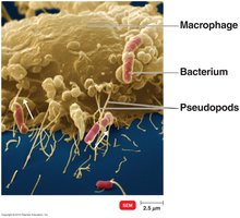
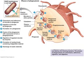
Phagocytosis
Phagocytosis is the ingestion and destruction of microbes by phagocytes such as neutrophils and macrophages.
Phagocytes: Cells that ingest and digest pathogens (e.g., neutrophils, macrophages).
Fixed Macrophages: Resident in specific tissues.
Wandering Macrophages: Move throughout tissues to sites of infection.
Steps of Phagocytosis
Chemotaxis: Chemical signals attract phagocytes to microbes.
Adherence: Phagocyte attaches to microbe surface.
Ingestion: Microbe is engulfed; opsonization (coating with serum proteins) enhances ingestion.
Digestion: Microbe is destroyed in a phagolysosome.
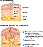
Inflammation
Inflammation is a local response to tissue damage, characterized by redness, swelling, pain, and heat. It aims to eliminate the initial cause of injury, clear out damaged cells, and establish tissue repair.
Acute-Phase Proteins: Activated during inflammation (e.g., complement, cytokines, kinins).
Vasodilation: Increases blood flow to the area (mediated by histamine, kinins, prostaglandins, leukotrienes).
Functions: Destroy injurious agents, limit effects, and repair tissue.
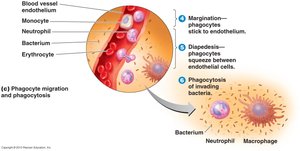
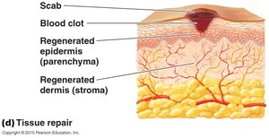
Phagocyte Migration and Tissue Repair
Margination: Phagocytes stick to blood vessel endothelium.
Diapedesis: Phagocytes squeeze between endothelial cells to reach infected tissue.
Tissue Repair: Stroma (supporting tissue) and parenchyma (functioning tissue) are regenerated after harmful substances are removed.
Fever
Fever is an abnormally high body temperature, often in response to infection. Cytokines trigger the hypothalamus to raise the body's temperature set point, which can inhibit pathogen growth and enhance immune responses.
Mechanism: Cytokines induce prostaglandin release, resetting the hypothalamus.
Effects: Vasoconstriction and shivering raise temperature; vasodilation and sweating lower it during recovery (crisis).
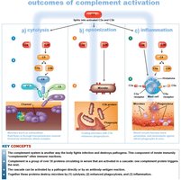
The Complement System
Overview and Activation Pathways
The complement system consists of serum proteins that enhance immune responses by promoting cytolysis, opsonization, and inflammation. Activation occurs via three pathways: classical, alternative, and lectin.
Classical Pathway: Triggered by antibodies binding to antigens, activating C1, then C2 and C4, leading to C3 activation.
Alternative Pathway: Initiated by C3 interacting with microbial surfaces and factors B, D, and P.
Lectin Pathway: Mannose-binding lectin (MBL) binds to microbial carbohydrates, activating C2 and C4, then C3.
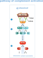
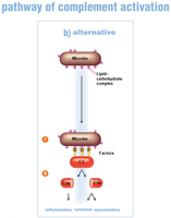
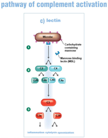
Outcomes of Complement Activation
Cytolysis: Formation of the membrane attack complex (MAC) that lyses pathogens.
Opsonization: Coating of microbes to enhance phagocytosis.
Inflammation: Complement proteins trigger histamine release from mast cells.
Regulation: Host regulatory proteins prevent excessive complement activation.
Clinical Relevance: Deficiencies in complement proteins increase susceptibility to infections; some pathogens evade complement by producing capsules.
Other Antimicrobial Substances
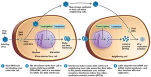
Interferons (IFNs)
Interferons are cytokines with antiviral properties. IFN-α and IFN-β are produced in response to viral infections and induce neighboring cells to produce antiviral proteins (AVPs). IFN-γ activates phagocytes to kill bacteria.
Iron-Binding Proteins
Iron-binding proteins (e.g., transferrin, lactoferrin, ferritin, hemoglobin) sequester iron, limiting its availability to microbes. Bacteria may produce siderophores to compete for iron.
Antimicrobial Peptides
Antimicrobial peptides are short proteins produced in response to microbial molecules. They inhibit cell wall synthesis, form pores in microbial membranes, and have broad-spectrum activity.
Additional info: These notes cover the main concepts of innate immunity, including physical and chemical barriers, cellular responses, inflammation, fever, the complement system, and antimicrobial substances, as outlined in Chapter 16 of a standard microbiology textbook.
