 Back
Back(Chapter 16) Innate Immunity: Nonspecific Defenses of the Host
Study Guide - Smart Notes
 Tailored notes based on your materials, expanded with key definitions, examples, and context.
Innate Immunity: Nonspecific Defenses of the Host
Overview of Immunity
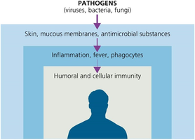
Immunity refers to the body's ability to resist and defend against infectious agents such as viruses, bacteria, and fungi. The immune system is divided into innate (nonspecific) and adaptive (specific) immunity. Innate immunity provides the first and second lines of defense, acting rapidly and broadly against pathogens without prior exposure.
First Line of Defense: Physical Factors
Skin
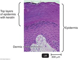
The skin acts as a formidable physical barrier to microbial invasion. It consists of two main layers:
Dermis: The inner layer, composed of connective tissue, provides structural support.
Epidermis: The outer layer, made of tightly packed epithelial cells containing keratin, a protective protein. The shedding and dryness of the skin inhibit microbial growth, and intact healthy epidermis is rarely penetrated by microbes.
Mucous Membranes
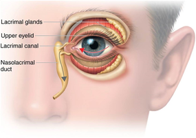
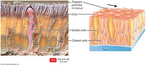
Mucous membranes line the gastrointestinal, respiratory, and genitourinary tracts. They consist of an epithelial layer that secretes mucus, a viscous glycoprotein that traps microbes and prevents the tracts from drying out. The lacrimal apparatus produces tears that wash the eye, helping to remove potential pathogens.
Other Physical Barriers
Ciliary escalator: Cilia in the respiratory tract move mucus and trapped microbes away from the lungs.
Epiglottis: Prevents microorganisms from entering the lower respiratory tract during swallowing.
Earwax: Blocks entry of microbes into the ear canal.
Urine and vaginal secretions: Flush out microbes from the urinary and vaginal tracts.
Peristalsis, defecation, vomiting, diarrhea: Expel pathogens from the gastrointestinal tract.
First Line of Defense: Chemical Factors
Chemical barriers enhance the effectiveness of physical barriers by creating hostile environments for pathogens:
Sebum: Forms a protective film and lowers the pH (3–5) of the skin, inhibiting microbial growth.
Lysozyme: An enzyme present in perspiration, tears, saliva, and urine that destroys bacterial cell walls.
Gastric juice: Has a low pH (1.2–3.0), destroying most bacteria and toxins in the stomach.
Vaginal secretions: Maintain a low pH (3–5), inhibiting microbial growth.
Normal Microbiota and Innate Immunity
Role of Normal Microbiota
Normal microbiota are the microorganisms that colonize the body without causing disease. They play a crucial role in innate immunity by:
Competing with pathogens for space and nutrients (microbial antagonism or competitive exclusion).
Producing substances harmful to pathogens.
Altering environmental conditions to inhibit pathogen survival.
Preventing the overgrowth of harmful microbes.
Contributing to the development of the immune system.
Commensalism describes a relationship where one organism benefits while the host is unharmed. Some normal microbiota can become opportunistic pathogens (e.g., Escherichia coli, Staphylococcus aureus).
Probiotics are live microbial cultures administered for beneficial effects, while prebiotics are nutrients that promote the growth of beneficial bacteria.
Second Line of Defense: Formed Elements in Blood
Blood Components
The second line of defense involves cellular components of blood, including:
Erythrocytes (red blood cells): Transport oxygen.
Leukocytes (white blood cells): Defend against pathogens.
Platelets: Involved in blood clotting.
All formed elements are produced in red bone marrow from stem cells through hematopoiesis.
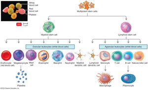
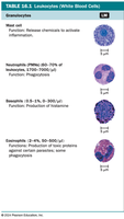
Granulocytes
Granulocytes are leukocytes with visible cytoplasmic granules:
Neutrophils: Highly phagocytic, first responders to infection.
Basophils: Release histamine, involved in allergic responses.
Eosinophils: Phagocytic, toxic to parasites and helminths.
Agranulocytes
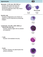
Agranulocytes are leukocytes with non-visible granules:
Monocytes: Mature into macrophages in tissues, where they become phagocytic.
Dendritic cells: Found in skin, mucous membranes, and thymus; initiate immune responses.
Lymphocytes: Include T cells, B cells, and natural killer (NK) cells; play roles in adaptive immunity.
Phagocytes and Phagocytosis
Types of Phagocytes
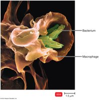
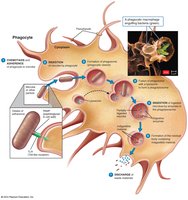
Phagocytosis is the process by which cells ingest and destroy microorganisms or debris. "Professional" phagocytes include neutrophils, eosinophils, and macrophages (derived from monocytes). Macrophages can be fixed (residing in tissues) or free (wandering in tissues).
Mechanism of Phagocytosis
The process of phagocytosis involves several steps:
Chemotaxis: Chemical signals attract phagocytes to the site of infection.
Adherence: Phagocyte attaches to the microbe, often enhanced by opsonization (coating with serum proteins such as antibodies or complement).
Ingestion: Pseudopods extend to engulf the microbe, forming a phagosome.
Digestion: Lysosomes fuse with the phagosome to form a phagolysosome, where enzymes and toxic oxygen products digest the microbe. Indigestible material is expelled by exocytosis.
Inflammation
Definition and Functions
Inflammation is a local defensive response to tissue damage caused by infection, physical, or chemical agents. The classic signs and symptoms are pain, redness, immobility, swelling (edema), and heat (PRISH). Functions include:
Destroying or limiting the effects of injurious agents.
Repairing and replacing damaged tissue.
Types and Stages of Inflammation
Acute inflammation: Rapid onset, short duration.
Chronic inflammation: Slow onset, long duration, potentially severe.
Stages include:
Vasodilation and increased vascular permeability
Phagocyte migration and phagocytosis
Tissue repair
Vasodilation and Increased Permeability
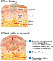
Vasodilation increases blood flow, causing redness and heat. Increased permeability allows defensive substances to reach the injured area, causing swelling. Chemicals such as histamine, kinins, prostaglandins, and cytokines mediate these changes. Blood clotting elements and pus (dead cells and fluids) may accumulate at the site.
Phagocyte Migration and Phagocytosis
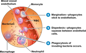
Phagocytes (mainly neutrophils and monocytes) migrate to the site of injury. Margination is the adherence of phagocytes to blood vessel walls, followed by diapedesis (movement through vessel walls). Monocytes mature into macrophages to clean up debris.
Tissue Repair
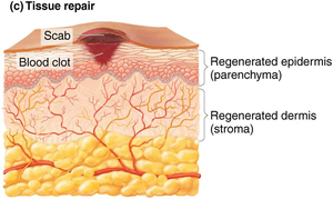
Tissue repair begins after harmful substances are removed. The stroma (supporting connective tissue) and parenchyma (functioning tissue) are regenerated to restore normal structure and function.
Fever
Fever is an abnormally high body temperature, often caused by infection. Cytokines stimulate the hypothalamus to raise the body's temperature set point. Fever enhances immune responses, inhibits pathogen growth, and speeds tissue repair, but can cause complications such as tachycardia, acidosis, dehydration, and, in severe cases, seizures or coma.
Antimicrobial Substances
Interferons
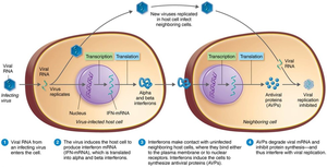
Interferons (IFNs) are cytokines produced by host cells in response to viral infection. They induce neighboring cells to produce antiviral proteins (AVPs) that inhibit viral replication. Types include alpha, beta, and gamma interferons, with gamma also activating phagocytes to kill bacteria.
Iron-Binding Proteins
Iron is essential for microbial growth. Host proteins such as transferrin, lactoferrin, ferritin, and hemoglobin tightly bind iron, limiting its availability to pathogens. Some bacteria produce siderophores to compete for iron.
Antimicrobial Peptides
Antimicrobial peptides are short proteins produced in response to microbial molecules. They have broad-spectrum activity, inhibiting cell wall synthesis, forming pores in membranes, and destroying nucleic acids. Examples include dermcidin, defensins, cathelicidins, and thrombocidin.
