 Back
BackInnate Immunity: Structure, Function, and Mechanisms
Study Guide - Smart Notes
 Tailored notes based on your materials, expanded with key definitions, examples, and context.
Innate Immunity
Concept of Immunity
Immunity refers to the body's ability to defend itself against disease-causing organisms and substances. It is divided into two main types: innate immunity and adaptive immunity.
Innate immunity: Defenses present at birth, always available, rapid response, non-specific, no memory response.
Adaptive immunity: Specific response to particular microbes, slower to respond, involves memory, mediated by lymphocytes (T cells and B cells).
Innate immunity is activated by protein receptors in the plasma membranes of defensive cells, such as Toll-like receptors (TLRs), which recognize pathogen-associated molecular patterns (PAMPs) like LPS, flagellin, peptidoglycan, and viral nucleic acids.
Defensive cells involved include macrophages and dendritic cells. Upon encountering PAMPs, TLRs induce these cells to release cytokines, which regulate immune responses and recruit other immune cells.
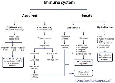
First Line of Defense: Physical Factors
The first line of defense consists of physical barriers that prevent the entry of pathogens.
Skin: Periodic shedding removes microbes; dryness inhibits growth; moist areas are more susceptible to infection.
Mucous membranes: Line GI, respiratory, and genitourinary tracts; mucus traps microbes; cilia propel trapped particles out.
Lacrimal apparatus: Produces tears, washing away microbes from the eye.
Saliva: Dilutes and washes microbes from the mouth.
Earwax: Prevents entry of microbes and other particles.
Urine flow: Cleanses the urethra.
Vaginal secretions: Expel microbes.
Peristalsis, defecation, vomiting, diarrhea: Expel microbes from the body.
First Line of Defense: Chemical Factors
Chemical barriers complement physical defenses by inhibiting microbial growth.
Sebum: Contains unsaturated fatty acids, inhibits bacteria and fungi.
Low skin pH: pH 3-5, inhibits microbial growth.
Sweat: Contains lysozyme, breaks down bacterial cell walls.
Earwax: Low pH due to fatty acids.
Saliva: Contains lysozyme, urea, uric acid, and immunoglobulin A (IgA).
Gastric juice: Highly acidic (pH 1.2-3.0), destroys bacteria and toxins.
Vaginal secretions: Acidic pH (3-5) due to lactic acid from Lactobacillus acidophilus.
Urine: Acidic pH (~6), contains lysozyme.
Normal Microbiota and Innate Immunity
Normal microbiota play a crucial role in innate immunity through microbial antagonism, competing with pathogens for nutrients and producing harmful substances. Probiotics are beneficial live cultures, while prebiotics promote growth of beneficial bacteria.
Second Line of Defense
Defensive Cells and Formed Elements in Blood
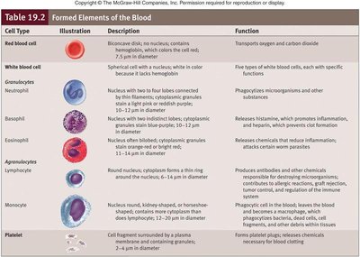
The second line of defense includes defensive cells, inflammation, fever, and antimicrobial substances. Blood contains plasma and formed elements: erythrocytes, leukocytes, and platelets. Leukocytes are divided into granulocytes and agranulocytes.
Granulocytes
Neutrophils: Highly phagocytic, active in initial infection stages.
Basophils: Release histamine, important in inflammation and allergy.
Eosinophils: Produce toxic proteins against parasites, somewhat phagocytic.
Agranulocytes
Monocytes: Mature into macrophages, phagocytic.
Dendritic cells: Destroy microbes by phagocytosis, initiate adaptive immunity.
Lymphocytes: Include natural killer (NK) cells, T cells, and B cells.
NK cells kill infected and tumor cells by releasing perforin and granzymes to induce apoptosis.
Leukocyte Response to Infection
During infection, leukocyte count increases (leukocytosis), while some diseases cause a decrease (leukopenia).
Lymphatic System
The lymphatic system includes lymph, vessels, and organs containing lymphoid tissue, protecting against ingested or inhaled microbes.
Phagocytes and Phagocytosis
Phagocytosis is the ingestion of microbes by white blood cells. Neutrophils dominate early infection, followed by macrophages. The process involves chemotaxis, adherence, ingestion, and digestion.
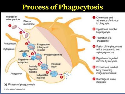
Chemotaxis: Attraction of phagocytes to microbes.
Adherence: Attachment facilitated by PAMPs and opsonization.
Ingestion: Pseudopods engulf microbe, forming a phagosome.
Digestion: Phagosome fuses with lysosome (phagolysosome), contents digested.
Some pathogens evade phagocytosis by inhibiting adherence, secreting toxins, surviving inside phagocytes, or forming biofilms.
Inflammation
Inflammation is a host response to tissue damage, characterized by redness, pain, heat, swelling, and sometimes loss of function. It can be acute or chronic, depending on the cause.
Functions: Destroy injurious agents, limit effects, repair tissue.
Stages: Vasodilation and increased permeability, phagocyte migration and phagocytosis, tissue repair.
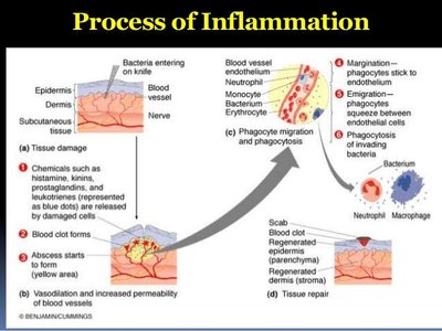
Chemicals involved include histamines, kinins, prostaglandins, and leukotrienes.
Fever
Fever is an abnormally high body temperature, usually caused by infection. The hypothalamus controls body temperature, which is raised by cytokines and prostaglandins during infection. Fever enhances immune responses, increases T cell production, intensifies interferon effects, and speeds up tissue repair.
Antibacterial Substances
Complement System
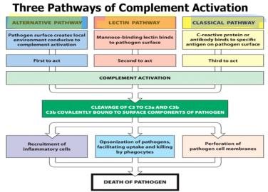
The complement system consists of over 30 proteins that destroy microbes via a cascade mechanism. Activation of C3 is crucial, leading to cytolysis, inflammation, and phagocytosis. There are three pathways:
Classical pathway: Initiated by antibodies binding to antigens.
Alternative pathway: Activated by complement proteins contacting pathogens.
Lectin pathway: Triggered by lectins binding to microbial carbohydrates.
Interferons
Interferons (IFNs) are antiviral proteins produced by lymphocytes and macrophages. Types include IFN-α, IFN-β (produced by virus-infected cells), and IFN-γ (produced by lymphocytes, induces neutrophils and macrophages to kill bacteria). Interferons are effective for short periods and play major roles in acute infections.
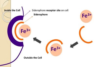
Iron-Binding Proteins
Pathogenic bacteria require iron for growth, but most iron in the body is bound to proteins (transferrin, lactoferrin, ferritin, hemoglobin). Bacteria secrete siderophores to compete for iron.
Antibacterial Peptides (AMPs)
AMPs are short peptides (12–50 amino acids) with broad antimicrobial activity. They inhibit cell wall synthesis, form membrane pores, and destroy DNA/RNA. Examples include dermcidin, defensins, cathelicidins, and thrombocidin.
Summary Table: Formed Elements of the Blood
Cell Type | Description | Function |
|---|---|---|
Red blood cell | Biconcave disk, no nucleus, contains hemoglobin | Transports oxygen and carbon dioxide |
Neutrophil | Spherical cell with nucleus, pale cytoplasm | Phagocytizes microorganisms and other substances |
Basophil | Nucleus with two lobes, cytoplasmic granules | Releases histamine, promotes inflammation |
Eosinophil | Nucleus with two lobes, cytoplasmic granules | Releases chemicals to reduce inflammation, attacks worm parasites |
Lymphocyte | Round nucleus, cytoplasm forms a thin ring | Produces antibodies and other chemicals, regulates immune response |
Monocyte | Nucleus is round, kidney-shaped, or horseshoe-shaped | Phagocytic cell in blood, becomes macrophage in tissues |
Platelet | Cell fragment surrounded by plasma membrane | Forms platelet plugs, releases chemicals for blood clotting |
