 Back
BackLectures 10, 11
Study Guide - Smart Notes
 Tailored notes based on your materials, expanded with key definitions, examples, and context.
Microbial Cell Structure
Overview of Cell Components
Microbial cells are the fundamental units of life, composed of various structures that perform essential functions. The cell envelope and cytoplasmic contents together define the cell's architecture and its ability to interact with the environment.
Envelope: Includes pili, outer membrane, capsule, wall, periplasm, cell membrane, and flagella.
Cytoplasmic contents: Nucleoid, cytoplasm, polysomes, vesicles.
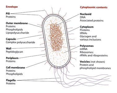
Cell Compartments and Functions
Each compartment within a microbial cell has a distinct role, contributing to the cell's survival, replication, and adaptation.
Cell membrane: Acts as a selective barrier, anchors proteins, and facilitates transport.
Nucleoid: Contains DNA and associated proteins, highly compacted for efficient storage.
Cytoplasm: Site of metabolic activity, contains proteins, tRNAs, glycogen, and inclusions.
Polysomes: Clusters of ribosomes translating mRNA.
Vesicles: Involved in protein and phospholipid transport (not always visible).
Microbial Diversity: Shapes and Sizes
Diversity of Cell Shapes
Microbes exhibit a wide range of shapes, each adapted to specific environmental challenges and functions.
Coccus: Spherical shape.
Rod: Cylindrical shape.
Spirillum: Spiral shape.
Spirochete, Hypha, Stalk, Filamentous: Specialized forms for motility, attachment, or dispersal.
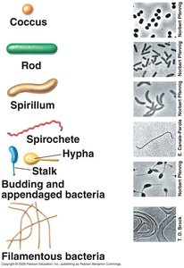
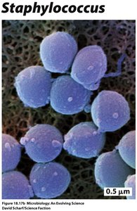
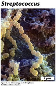
Examples of Microbial Shapes
Specific genera demonstrate characteristic shapes, which can be observed under the microscope.
Staphylococcus: Clustered cocci.
Streptococcus: Chains of cocci.
Archaeal Cell Structures
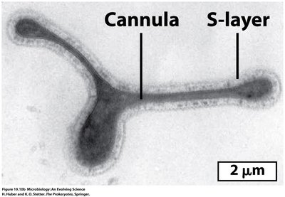
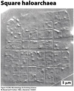
Archaea display unique cell shapes and surface structures, such as cannulae and S-layers, and some have square morphologies.
Cannula: Tubular structures connecting cells.
S-layer: Surface protein layer providing structural support.
Square haloarchaea: Unique square-shaped cells found in extreme environments.
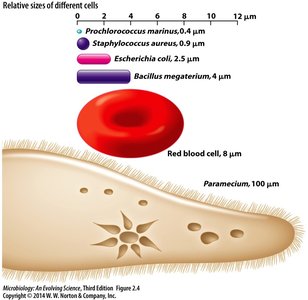
Microbial Cell Sizes
Microbes vary greatly in size, from tiny bacteria to larger eukaryotic cells.
Prochlorococcus marinus: 0.4 μm
Staphylococcus aureus: 0.9 μm
Escherichia coli: 2.5 μm
Bacillus megaterium: 4 μm
Red blood cell: 8 μm
Paramecium: 100 μm
Cell Composition: Monomers and Macromolecules
Monomers: Building Blocks of Cells
Cells are constructed from four basic types of monomers: sugars, amino acids, purines and pyrimidines, and lipids. These monomers are assembled into macromolecules essential for cellular function.
Sugars (carbohydrates): Serve as carbon and energy sources, storage materials, adhesives, and structural components.
Amino acids: Form proteins via peptide bonds.
Purines and pyrimidines: Build nucleic acids (DNA and RNA).
Lipids: Compose membranes and energy reserves.
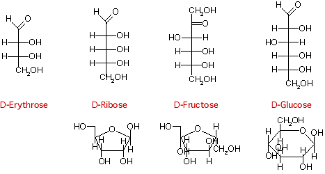
Sugar Structures and Functions
Sugars typically contain 3 to 7 carbons and exist in open or closed forms. Closed forms are classified as furanosides (five atoms) or pyranosides (six atoms).
Aldoses: Sugars with an aldehyde group at C-1.
Ketoses: Sugars with a ketone group at C-2.

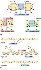
Sugar Polymers
Sugars are polymerized to form polysaccharides, which serve as energy storage (e.g., starch, glycogen) or structural materials (e.g., cellulose).
Glycosidic bonds: Link monosaccharides in polysaccharides.
Starch and glycogen: Contain α-1,4 and α-1,6 bonds.
Cellulose: Contains β-1,4 bonds.
Proteins and Amino Acids
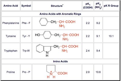
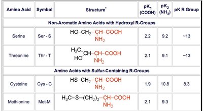
Amino Acids: Structure and Properties
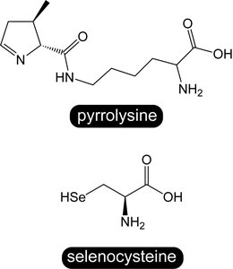
Amino acids are organic acids with an amino group at the alpha carbon. They are distinguished by their side chains and are linked by peptide bonds to form proteins. There are 22 proteinogenic amino acids, including rare ones like pyrrolysine and selenocysteine.
pKa: The pH at which ionizable groups are present at equal concentrations of protonated and deprotonated forms.
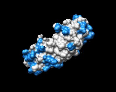
Protein Structure
Proteins have four levels of structure:
Primary: Sequence of amino acids.
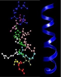
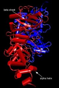
Secondary: Localized folding (α helix, β sheet).
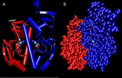
Tertiary: Overall folding stabilized by hydrophobic interactions, hydrogen bonds, ionic interactions, and disulfide bonds.
Quaternary: Assembly of multiple polypeptides.
Nucleic Acids: DNA and RNA
Nucleotides and Nucleic Acid Structure
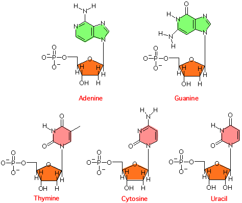
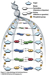
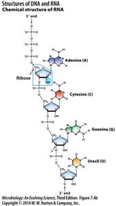
Nucleic acids are polymers of nucleotides, which consist of a sugar, phosphate group, and nitrogenous base. DNA and RNA differ in their sugar and base composition.
DNA: Contains deoxyribose and bases adenine, guanine, cytosine, thymine.
RNA: Contains ribose and bases adenine, guanine, cytosine, uracil.
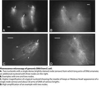
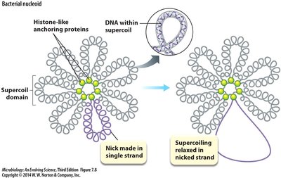
Nucleoid: DNA Compaction in Prokaryotes
The nucleoid is a highly compacted DNA-protein complex in prokaryotes. DNA is organized into loops or domains, anchored by histone-like proteins, allowing efficient storage and accessibility for replication and transcription.
Compaction: E. coli genome is compacted ~1000X to fit within the cell.
Organization: DNA binding proteins bend and condense DNA.
Genome Size and Function
Prokaryotic genomes vary in size and complexity, influencing the cell's metabolic capabilities and adaptability.
Nucleic Acid Function
DNA: Storage of hereditary information.
RNA: Translation (mRNA, tRNA, rRNA).
Ribosomes: Protein synthesis machines, composed of RNA and protein.
Lipids and Membranes
Lipid Structure and Function
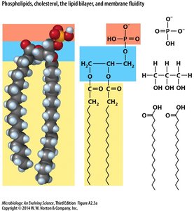
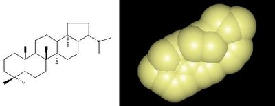
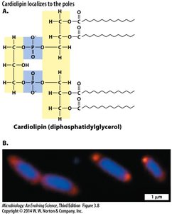
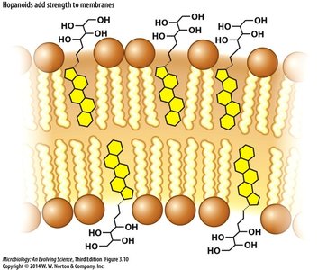
Lipids are essential for membrane structure and function. In microbes, phospholipids are predominant, with variations in fatty acid chains and head groups. Some bacteria contain hopanoids, which add rigidity to membranes.
Phospholipids: Glycerol backbone, two fatty acyl esters (hydrophobic), phosphate group (hydrophilic).
Hopanoids: Planar, rigid molecules that strengthen membranes.
Membrane diversity: Over 100 lipid structures known, with variation in charge, melting point, and shape.

Archaeal Membrane Lipids
Archaeal membranes differ from bacterial ones, often forming lipid monolayers instead of bilayers, and using unique ether-linked lipids.
Membrane Structure and Function
The cell membrane is a fluid mosaic, stabilized by hydrophobic interactions, hydrogen bonds, and ionic interactions. It acts as a selective barrier, anchors proteins, and facilitates transport.
Passive transport: Diffusion, facilitated diffusion.
Active transport: Ion-coupled, ATP-binding cassette (ABC) transporters, group translocation.
Small Molecules in Cells
Small Molecule Functions
Small molecules, such as coenzymes, cofactors, and ions, are vital for cellular metabolism and energy conservation.
NAD: Universal electron carrier.
Summary Table: Cell Building Blocks
Building Block | Function | Example |
|---|---|---|
Sugars | Energy, storage, structure | Glucose, glycogen, cellulose |
Amino acids | Protein synthesis | Serine, tyrosine, tryptophan |
Pyrimidines/Purines | Nucleic acid synthesis | Adenine, guanine, cytosine, thymine, uracil |
Lipids | Membrane structure, energy storage | Phospholipids, hopanoids |
Key Equations
pKa Definition:
Peptide Bond Formation:
DNA Double Helix Base Pairing:
Additional info: Academic context was added to clarify the functions and structures of cell components, monomers, and macromolecules, as well as to provide examples and equations relevant to microbiology students.
