 Back
Microbiology Study Guide: Antimicrobial Agents, Viruses, and Infectious Diseases
Study Guide - Smart Notes
 Tailored notes based on your materials, expanded with key definitions, examples, and context.
Controlling Microbial Growth in the Body: Antimicrobial Agents
History and Types of Antimicrobial Agents
Antimicrobial agents are chemotherapeutic drugs used to treat diseases caused by microorganisms. The development of these agents began in the early 20th century, with key discoveries by Paul Ehrlich ("magic bullets" and chemotherapy), Alexander Fleming (penicillin), and Gerhard Domagk (sulfanilamide).
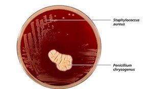
Antibiotics: Naturally produced by organisms, e.g., Penicillium secreting penicillin.
Semisynthetic drugs: Chemically modified antibiotics.
Synthetic drugs: Fully synthesized in laboratories.
Mechanisms of Antimicrobial Action
Antimicrobial drugs act through selective toxicity, targeting pathogens more than host cells. The main mechanisms include:
Inhibition of Cell Wall Synthesis: Interferes with peptidoglycan formation, weakening bacterial cell walls. Most effective against actively growing bacteria with thick peptidoglycan layers (e.g., penicillin, vancomycin).
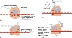
Inhibition of Protein Synthesis: Targets differences in ribosome structure between prokaryotes and eukaryotes. Drugs like tetracycline and chloramphenicol inhibit prokaryotic ribosomes but may affect mitochondrial ribosomes in eukaryotes.
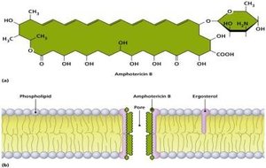
Disruption of Cytoplasmic Membranes: Drugs like Amphotericin B bind to ergosterol in fungal membranes, forming pores and causing cell lysis. Human membranes can also be affected.
Inhibition of Metabolic Pathways: Structural analogs (e.g., sulfonamides) block essential metabolic reactions by mimicking substrates like PABA.
Inhibition of Nucleic Acid Synthesis: Drugs such as quinolones and rifampin target DNA or RNA synthesis, with varying specificity for prokaryotes and eukaryotes.
Prevention of Virus Attachment: Attachment antagonists block viral entry, still under development.
Clinical Considerations in Prescribing Antimicrobials
The ideal antimicrobial agent is readily available, inexpensive, stable, easily administered, nontoxic, and selectively toxic. Since no drug is perfect, clinicians consider spectrum of action, organism sensitivity, administration route, and safety/side effects.
Spectrum of Action
Narrow-spectrum drugs: Target specific pathogens, minimizing impact on normal flora (e.g., penicillin).
Broad-spectrum drugs: Affect a wide range of pathogens but may disrupt normal microbiota, leading to opportunistic infections.
Efficacy Testing
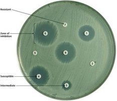
Diffusion Susceptibility Test (Kirby-Bauer): Measures zones of inhibition around drug-impregnated discs on agar plates to assess sensitivity.
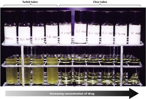
Minimum Inhibitory Concentration (MIC): Lowest drug concentration preventing visible growth in broth dilution tests.
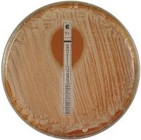
Etest: Combines MIC and diffusion test using a drug gradient strip.
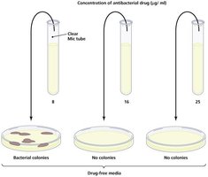
Minimum Bactericidal Concentration (MBC): Lowest concentration that kills bacteria, determined by subculturing from MIC tubes.
Routes of Administration
Topical: For surface infections.
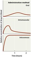
Oral: Simple but may result in lower drug concentration at infection site.
Intramuscular (IM): Faster delivery, moderate concentration.
Intravenous (IV): Fastest and highest concentration.
Safety and Side Effects
Toxicity: Adverse effects on host organs or fetus.
Allergies: Immune reactions, including anaphylactic shock.
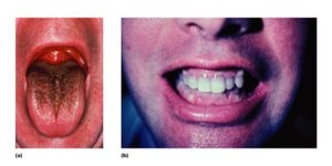
Disruption of Normal Microbiota: Can lead to superinfections by opportunistic pathogens.
Resistance to Antimicrobial Drugs
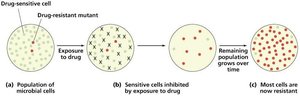
Pathogens develop resistance through mutations and horizontal gene transfer (R-plasmids). Resistance mechanisms include enzyme production, altered drug targets, reduced drug entry, and drug efflux.
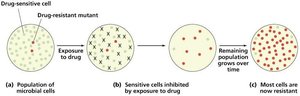
Retarding Resistance
Use drugs at high enough concentrations and for sufficient duration.
Combine drugs for synergistic effects.
Limit use to necessary cases.
Develop new drug generations and search for novel agents.
Characterizing and Classifying Viruses, Viroids, and Prions
Characteristics of Viruses
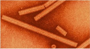
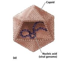
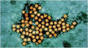
Viruses are acellular, obligate intracellular parasites composed of DNA or RNA, lacking membranes and organelles. Outside cells, they exist as virions with a protein capsid surrounding the nucleic acid core.
Genetic Material
Genome can be dsDNA, ssDNA, dsRNA, or ssRNA.
Usually smaller than host genome.
Hosts and Specificity
Viruses infect specific cell types and hosts (e.g., HIV infects human T lymphocytes).
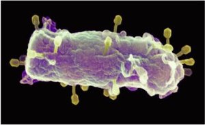
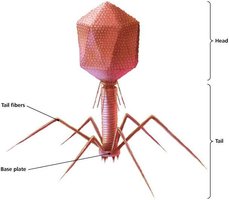
Bacteriophages infect bacteria; plant viruses infect plants.
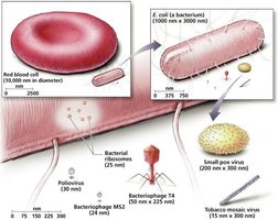
Sizes of Viruses
Viruses are much smaller than bacteria and human cells.
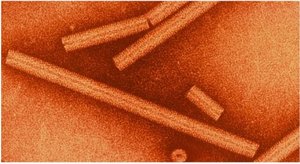
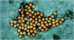
Capsid Morphology and Viral Shapes
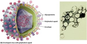
Capsids are made of capsomeres and provide protection and attachment.
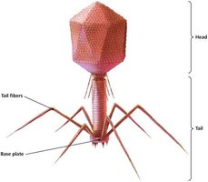
Shapes: helical, polyhedral, and complex.
The Viral Envelope
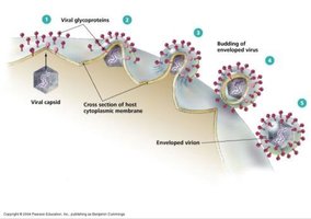
Enveloped virions have a phospholipid membrane with embedded proteins (spikes).
Envelope acquired during release from host cell.
Classification of Viruses
Based on genetic material, size, shape, and envelope presence.
Viral Replication
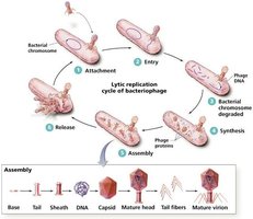
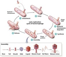
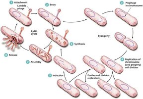
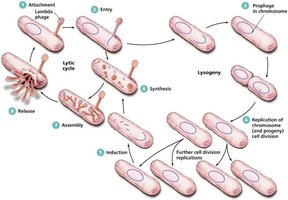
Lytic Replication of Bacteriophages
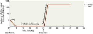
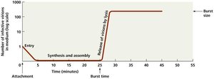
Stages: attachment, entry, synthesis, assembly, release.
Burst time: time from attachment to release.
Burst number: number of virions released per cell.
Lysogeny
Lysogenic phages remain inactive as prophages in host DNA.
Lysogenic conversion can increase bacterial virulence.
Induction triggers lytic cycle.
Replication of Animal Viruses
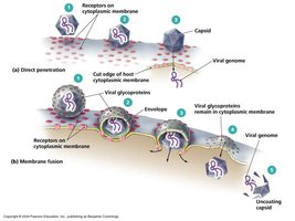
Attachment via glycoprotein spikes.
Entry by membrane fusion, phagocytosis, or direct penetration.
Synthesis depends on genome type (dsDNA, ssDNA, +ssRNA, -ssRNA, retrovirus).
Assembly in nucleus (DNA viruses) or cytosol (RNA viruses).
Budding releases enveloped viruses; naked viruses released by lysis or exocytosis.
Latency of Animal Viruses
Latent (proviruses) may remain dormant for years; some integrate into host genome.
The Role of Viruses in Cancer
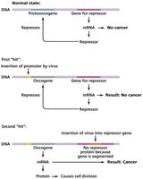
Oncogenes and tumor repressor genes regulate cell division.
Viruses can activate oncogenes, causing neoplasia and cancer.
Culturing Viruses in the Laboratory
Viruses are grown in whole organisms, embryonated eggs, or cell cultures.
Other Parasitic Particles: Viroids and Prions
Viroids
Infectious RNA molecules without capsids, infect plants.
Prions
Proteinaceous infectious agents; cause normal PrP to misfold into beta-pleated sheets.
Prion diseases (spongiform encephalopathies) are fatal and resistant to sterilization.
Infection, Infectious Diseases, and Epidemiology
Symbiotic Relationships Between Microbes and Hosts
Mutualism: Both benefit (e.g., E. coli in colon).
Commensalism: One benefits, other unaffected.
Parasitism: One benefits, other harmed (pathogens).
Normal Microbiota
Resident microbiota: Permanent, location-specific.
Transient microbiota: Temporary, replaced by competition.
Acquired at birth and throughout life.
Opportunistic Pathogens
Normal flora can cause disease if environment changes or immune system is compromised.
Microbial antagonism keeps opportunists in check.
Reservoirs of Infectious Diseases
Animal reservoirs: Zoonoses transmitted from animals to humans.
Human carriers: Asymptomatic or symptomatic individuals.
Nonliving reservoirs: Soil, water, food.
Movement of Microbes into Hosts: Infection
Portals of Entry
Skin, mucous membranes, placenta, parenteral routes.
Mucous membranes (respiratory, GI, urinary, reproductive tracts) are main entry points.
The Role of Adhesion in Infection
Adhesion factors (ligands) enable pathogens to attach to host cells.
Biofilms are communities of attached organisms (e.g., dental plaque).
The Nature of Infectious Diseases
Infection: Successful colonization.
Disease: Change in host health (morbidity).
Manifestations: Symptoms, Signs, Syndromes
Signs: Objective, measurable (fever, rash).
Symptoms: Subjective (pain, dizziness).
Syndrome: Set of signs and symptoms for a disease.
Causation: Etiology and Koch’s Postulates
Koch’s postulates identify causative agents of disease.
Virulence Factors of Infectious Disease
Pathogenicity: Ability to cause disease.
Virulence: Degree of pathogenicity.
Virulence factors: adhesion, biofilms, extracellular enzymes, toxins, antiphagocytic factors.
Extracellular Enzymes
Hyaluronidase, collagenase, coagulase, kinases aid invasion and evasion.
Toxins
Exotoxins: Secreted, destroy host cells or metabolism (cytotoxins, neurotoxins, enterotoxins).
Endotoxins: Released from cell wall (e.g., Lipid A in Gram-negative bacteria).
Antitoxins and toxoids provide immunity.
Antiphagocytic Factors
Capsules, M proteins, leukocidins prevent phagocytosis.
The Stages of Infectious Disease
Incubation: Time from infection to symptoms.
Prodromal: Mild symptoms appear.
Illness: Severe symptoms, immune response not fully active.
Decline: Symptoms decrease, recovery begins.
Convalescence: Repair and return to normal health.
Portals of Exit
Pathogens exit via body fluids (saliva, mucus, blood).
Modes of Infectious Disease Transmission
Contact transmission: Direct, indirect (fomites), droplet.
Vehicle transmission: Airborne, waterborne, foodborne, body fluids.
Vector transmission: Biological (host and carrier), mechanical (carrier only).
Classification of Infectious Diseases
By longevity/severity: Acute, chronic, latent, subacute.
By transmission: Communicable, contagious, non-communicable.
Epidemiology of Infectious Diseases
Incidence: New cases in a population/time.
Prevalence: Total cases in a population/time.
Endemic: Constant presence.
Sporadic: Occasional cases.
Epidemic: Increased occurrence.
Pandemic: Occurrence across continents.
Epidemiological Studies
Descriptive: Data collection and case tracking.
Analytical: Cause, transmission, prevention analysis.
Experimental: Hypothesis testing.
Hospital Epidemiology: Nosocomial Infections
Exogenous: From health care environment.
Endogenous: From patient’s own microbiota.
Iatrogenic: From medical procedures.
Superinfections: From antibiotic misuse.
Control of Nosocomial Infections
Use of sterile technique, hand washing, proper disposal of sharps, and public health education.
Epidemiology and Public Health
Public health agencies collect, analyze, and share data to prevent and control disease outbreaks.
Sharing of Data
Local, state, federal (CDC), and international (WHO) agencies collaborate.
Public Health Education
Agencies educate the public on disease prevention and response.
