 Back
BackPathogenic DNA Viruses: Herpesviridae and Related Human Diseases
Study Guide - Smart Notes
 Tailored notes based on your materials, expanded with key definitions, examples, and context.
Pathogenic DNA Viruses
Overview of DNA Virus Families
Pathogenic DNA viruses are classified into seven families based on their genetic material, structure, and disease associations. Understanding these families is essential for recognizing their clinical significance and epidemiology.
Poxviridae: Double-stranded, enveloped, complex capsid; causes smallpox, cowpox, molluscum contagiosum.
Herpesviridae: Double-stranded, enveloped, icosahedral capsid; includes HHV-1 to HHV-8, causing a range of diseases from cold sores to cancers.
Papillomaviridae: Double-stranded, naked, icosahedral capsid; causes warts and cancers.
Polyomaviridae: Double-stranded, naked, icosahedral capsid; causes progressive multifocal leukoencephalopathy.
Other families: Adenoviridae, Hepadnaviridae, Parvoviridae (not detailed in these notes).
Family | Strand Type | Envelope | Capsid Symmetry | Size (nm) | Representative Genera (Disease) |
|---|---|---|---|---|---|
Poxviridae | Double | Enveloped | Complex | 200–300 | Orthopoxvirus (smallpox, cowpox), Molluscipoxvirus (molluscum contagiosum) |
Herpesviridae | Double | Enveloped | Icosahedral | 150–200 | HHV-1, HHV-2, Varicellovirus, Epstein-Barr virus, Cytomegalovirus, Roseolovirus |
Papillomaviridae | Double | Naked | Icosahedral | 45–55 | Papillomavirus (warts, cancers) |
Polyomaviridae | Double | Naked | Icosahedral | 45–55 | Polyomavirus (leukoencephalopathy) |
Herpesviridae: General Characteristics
The Herpesviridae family includes linear double-stranded DNA viruses with enveloped icosahedral capsids. They are the most prevalent DNA viruses in humans and are notable for their ability to establish latency within host cells, often reactivating to cause recurrent disease.
Envelope: Acquired from host cell's nuclear membrane.
Latency: Viruses remain inactive in infected cells, often in neural ganglia.
Species Naming: "HHV" followed by a number (e.g., HHV-1, HHV-2).
Human Herpesvirus 1 and 2 (HHV-1, HHV-2)
HHV-1 and HHV-2, formerly known as herpes simplex viruses (HSV), are responsible for a variety of slow-spreading skin and mucosal lesions. After primary infection, these viruses establish latency in neural ganglia and may reactivate later in life.
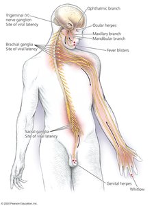
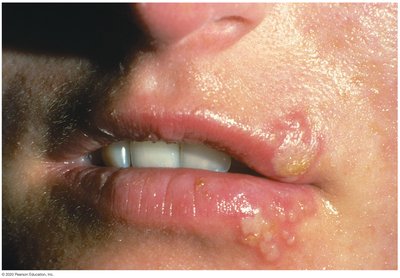
HHV-1: Primarily causes oral herpes (fever blisters, cold sores), but can also cause genital herpes, ocular herpes, whitlow, and neonatal herpes.
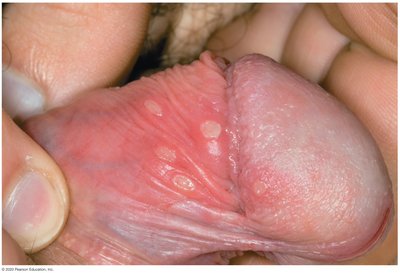
HHV-2: Primarily causes genital herpes, but can also cause oral lesions, whitlow, and neonatal herpes.
Types of HHV-1 and HHV-2 Infections
Oral Herpes: HHV-1 causes fever blisters/cold sores; can extend to gingivostomatitis and pharyngitis.
Genital Herpes: HHV-2 causes most genital herpes; painful lesions on genitalia, can also cause oral lesions via oral sex.
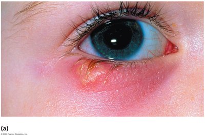
Ocular Herpes: Reactivation of HHV-1 in the eye; symptoms include gritty feeling, conjunctivitis, pain, sensitivity to light, and corneal lesions. Recurrent infections may lead to blindness.
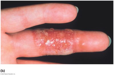
Whitlow: Inflamed blister on the finger, often seen in children and healthcare workers.
Neonatal Herpes: Severe infections in newborns, acquired in utero or during birth; high mortality rate.
Other: Herpes gladiatorum (athletes), encephalitis, meningitis, pneumonia in immunosuppressed individuals.
Comparative Epidemiology and Pathology of HHV-1 and HHV-2
HHV-1 (HSV-1) | HHV-2 (HSV-2) |
|---|---|
90% of cold sores/fever blisters; whitlow | 85% of genital herpes cases |
Close contact | Sexual intercourse |
Trigeminal and brachial ganglia (latency) | Sacral ganglia (latency) |
Face, mouth, rarely trunk | External genitalia, thighs, buttocks, anus |
Pharyngitis, gingivostomatitis, ocular herpes, gladiatorum | 30% neonatal herpes, 10% oral herpes |
Epidemiology and Pathogenesis
Transmission: Close body contact; active lesions are main source; asymptomatic carriers shed HHV-2 genitally.
Entry: Through cracks/cuts in mucous membranes.
Replication: In epithelial cells, leading to lesion formation.
Spread: Cell-to-cell via syncytia formation.
Age: HHV-1: casual contact in children; HHV-2: sexual activity (15–29 years).
Diagnosis, Treatment, and Prevention
Diagnosis: Characteristic lesions are often diagnostic.
Treatment: Valaciclovir for cold sores/genital herpes; iododeoxyuridine/trifluridine for ocular herpes; acyclovir during pregnancy.
Prevention: Glove use for healthcare workers; sexual abstinence or sex between uninfected partners.
Human Herpesvirus 3 (Varicella-Zoster Virus, HHV-3)
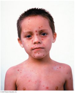
HHV-3 causes two distinct diseases: varicella (chickenpox) and herpes zoster (shingles). Chickenpox is common in children, while shingles typically affects adults.
Chickenpox: Highly infectious; virus enters via respiratory tract or eyes, spreads through blood, causes characteristic skin lesions after 2–3 weeks.
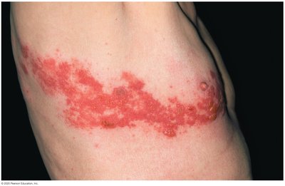
Shingles: Reactivation of latent virus; rash appears along a dermatome.
Epidemiology and Pathogenesis
Chickenpox: Mild in children, more severe in adults.
Shingles: Latent virus reactivates, producing a painful rash.
Diagnosis, Treatment, and Prevention
Diagnosis: Chickenpox diagnosed by characteristic lesions; shingles lesions can be more difficult to diagnose.
Treatment: Chickenpox is usually self-limiting; shingles treatment focuses on symptom management.
Prevention: Vaccines available for both chickenpox and shingles; exposure difficult to prevent due to viral shedding before symptoms.
Human Herpesvirus 4 (Epstein-Barr Virus, HHV-4)
Epstein-Barr virus (EBV) is associated with several diseases, including infectious mononucleosis, Burkitt’s lymphoma, and Hodgkin’s lymphoma. It is transmitted primarily via saliva.
Initial Infection: Epithelium of pharynx and parotid glands.
Spread: Invades B lymphocytes, becomes latent, suppresses apoptosis.
Immune Response: Cytotoxic T cells kill infected B cells, causing mononucleosis symptoms.
Diagnosis, Treatment, and Prevention
Diagnosis: Based on characteristic signs.
Treatment: Chemotherapy for lymphomas; symptomatic relief for mono; no effective treatment for other conditions.
Prevention: Difficult due to widespread nature and saliva transmission.
Human Herpesvirus 5 (Cytomegalovirus, HHV-5)
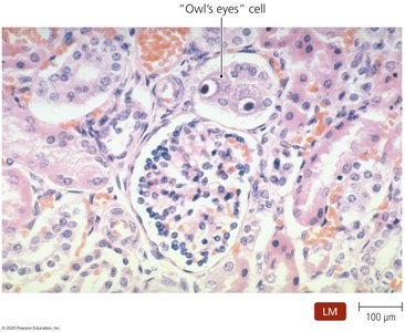
Cytomegalovirus (CMV) is a common human infection, often asymptomatic but can cause severe complications in fetuses, newborns, and immunodeficient patients. Infected cells become enlarged, known as "owl's eye" cells.
Transmission: Bodily secretions, sexual intercourse, in utero exposure, birth, blood transfusions, organ transplants.
Complications: Mental retardation, hearing/visual damage in newborns; pneumonia, blindness, mononucleosis in immunosuppressed adults.
Diagnosis, Treatment, and Prevention
Diagnosis: Detection of enlarged cells and cellular inclusions; ELISA or DNA probes.
Treatment: Difficult for fetuses/newborns; damage often occurs before detection; adult treatment is also challenging.
Prevention: Difficult due to widespread transmission.
Other Herpesvirus Infections
Other human herpesviruses include HHV-6 (Roseolovirus) and HHV-8 (Rhadinovirus).
HHV-6: Causes roseola (pink rash), may be linked to multiple sclerosis, can cause mononucleosis-like symptoms, may increase susceptibility to AIDS.
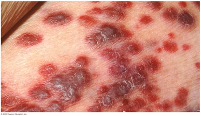
HHV-8: Causes Kaposi's sarcoma, a cancer often seen in AIDS patients.

Review Questions and Clinical Correlations
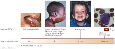
EBV and Immune Response: Vigorous cellular immunity leads to infectious mononucleosis; poor immunity can result in Burkitt’s lymphoma or Hodgkin’s lymphoma.
Genital Herpes: Most likely caused by HHV-2.
Neutropenia: EBV infection can cause a lower than normal number of neutrophils in the blood.
Summary Table: Herpesvirus Diseases and Immunity
State of Cellular Immunity | Disease |
|---|---|
Lacking | Oral hairy leukoplakia |
Poor | Burkitt's lymphoma, nasopharyngeal cancer, chronic fatigue syndrome, Hodgkin's lymphoma |
Normal | Asymptomatic |
Vigorous | Infectious mononucleosis |
Additional info: These notes expand on brief points with academic context, including definitions, examples, and tables for clarity. Images are included only when directly relevant to the explanation of the paragraph.
