 Back
BackPrinciples of Disease, Pathogenesis, and Immunology: The Lymphatic System and Host Defenses
Study Guide - Smart Notes
 Tailored notes based on your materials, expanded with key definitions, examples, and context.
Principles of Disease and Epidemiology
Introduction to Health and Disease
Health is defined as the ability to adapt and self-manage in the face of physical, mental, and social challenges. Disease represents an impaired ability to maintain homeostasis, often due to pathogenic microorganisms or other factors.
Pathology: The scientific study of disease.
Etiology: The cause of a disease.
Pathogenesis: The development of disease and the chain of events leading to that disease.
Pathophysiology: The functional changes associated with or resulting from disease.
Pathogenicity: The ability of a microorganism to cause disease.
Virulence: The degree of pathogenicity.
Microbiome and Host Relationships
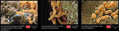
The human body is colonized by a vast array of microorganisms, collectively known as the microbiota. These organisms can be permanent residents (normal flora) or transient.
Colonization: Establishment of microbial residence without causing disease.
Normal Microbiota: Microorganisms that colonize the body without causing disease under normal conditions.
Transient Microbiota: Microbes present for days, weeks, or months, then disappear.
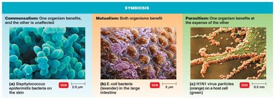
Types of Symbiotic Relationships
Microorganisms interact with their hosts in various ways, including:
Commensalism: One organism benefits, the other is unaffected (e.g., Staphylococcus epidermidis on skin).
Mutualism: Both organisms benefit (e.g., Escherichia coli in the large intestine synthesizes vitamins for the host).
Parasitism: One organism benefits at the expense of the other (e.g., viruses infecting host cells).
Microbial Antagonism and Opportunism
Normal flora can prevent the overgrowth of pathogens through microbial antagonism (competitive exclusion). Some microbes become pathogenic under certain conditions (opportunistic pathogens), such as changes in location, concentration, or host immunity.
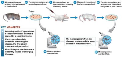
Etiology and Koch’s Postulates
Etiology is the study of disease causation. Koch’s postulates are a set of criteria used to establish a causative relationship between a microbe and a disease:
The microorganism must be found in all cases of the disease.
It must be isolated and grown in pure culture.
The cultured microorganism should cause disease when introduced into a healthy host.
It must be re-isolated from the experimentally infected host.
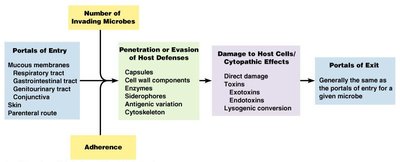
Portals of Entry, Adherence, and Pathogenesis
Pathogens enter the host through specific portals (e.g., mucous membranes, skin, parenteral route), adhere to host tissues, and evade or penetrate host defenses using various mechanisms (e.g., capsules, enzymes, antigenic variation).
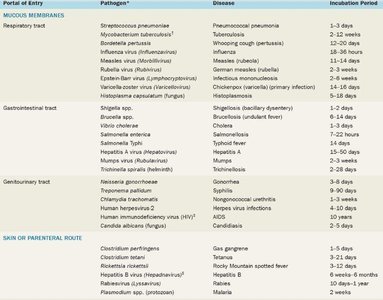
Portals of Entry and Incubation Periods
Different pathogens use specific portals of entry and have characteristic incubation periods before symptoms appear.
Portal of Entry | Pathogen | Disease | Incubation Period |
|---|---|---|---|
Respiratory tract | Streptococcus pneumoniae | Pneumococcal pneumonia | 1–3 days |
Gastrointestinal tract | Shigella spp. | Bacillary dysentery | 1–7 days |
Genitourinary tract | Neisseria gonorrhoeae | Gonorrhea | 2–7 days |
Skin/parenteral route | Clostridium perfringens | Gas gangrene | 1–5 days |
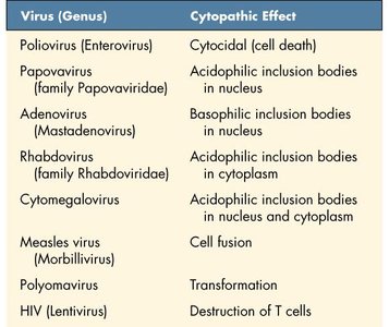
Cytopathic Effects of Viruses
Viruses can cause specific cytopathic effects (CPE) in host cells, such as cell death, inclusion bodies, cell fusion, transformation, or immune cell destruction.
Virus (Genus) | Cytopathic Effect |
|---|---|
Poliovirus (Enterovirus) | Cytocidal (cell death) |
Measles virus (Morbillivirus) | Cell fusion |
HIV (Lentivirus) | Destruction of T cells |
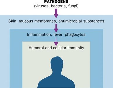
Host Defenses and Immunology
Overview of Host Defenses
The human body employs multiple layers of defense against pathogens:
First line: Physical and chemical barriers (skin, mucous membranes, antimicrobial substances).
Second line: Inflammation, fever, phagocytes.
Third line: Adaptive (humoral and cellular) immunity.
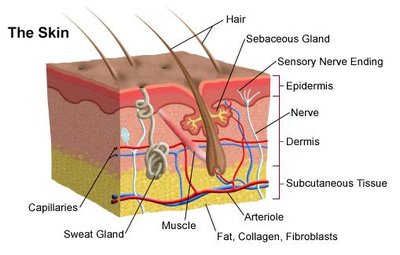
Physical Barriers: The Skin
The skin acts as a primary physical barrier, composed of multiple layers of cells and associated structures that prevent pathogen entry.
Epidermis: Outermost layer, tightly packed cells, includes the stratum corneum.
Dermis: Contains connective tissue, blood vessels, and immune cells.
Associated structures: Sweat glands (produce salt and antimicrobial peptides), sebaceous glands (produce sebum).
Mucosal Surfaces and Secretions
Mucous membranes line all body cavities exposed to the external environment and provide a barrier to pathogens. Secretions such as tears, saliva, and mucus contain antimicrobial substances like lysozyme.
Innate Immune Cells
White blood cells (leukocytes) are key players in innate immunity. They include granulocytes (basophils, eosinophils, neutrophils) and agranulocytes (lymphocytes, monocytes).
Cell Type | Description | Function |
|---|---|---|
Basophil | Granulocyte | Releases histamines that cause inflammation |
Eosinophil | Granulocyte | Kills parasites with oxidative burst |
Neutrophil | Both | Phagocytizes bacteria and fungi |
Monocyte | Agranulocyte | Precursor to macrophages |
Dendritic cell | Agranulocyte | Antigen-presenting cell |
Natural killer (NK) cell | Agranulocyte | Kills cancer and virus-infected cells |

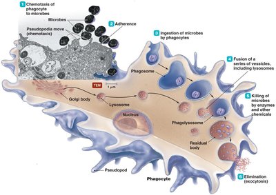
Phagocytosis
Phagocytosis is the process by which certain cells (e.g., neutrophils, macrophages) ingest and destroy pathogens. The stages include chemotaxis, adherence, ingestion, maturation (fusion with lysosomes), killing, and elimination.
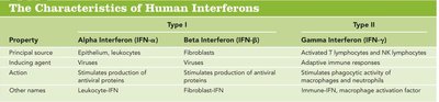
Interferons
Interferons are cytokines that play a crucial role in antiviral defense. They are classified as Type I (alpha, beta) and Type II (gamma), each with distinct sources and functions.
Type | Principal Source | Inducing Agent | Action |
|---|---|---|---|
Alpha (IFN-α) | Epithelium, leukocytes | Viruses | Stimulates production of antiviral proteins |
Beta (IFN-β) | Fibroblasts | Viruses | Stimulates production of antiviral proteins |
Gamma (IFN-γ) | Activated T lymphocytes, NK lymphocytes | Adaptive immunity | Stimulates phagocytic activity |
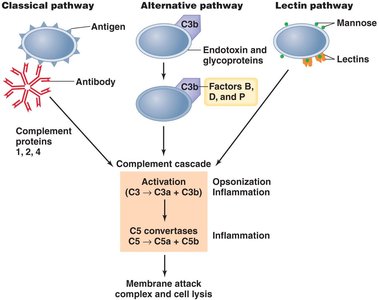
The Complement System
The complement system consists of serum proteins that enhance (complement) the ability of antibodies and phagocytic cells to clear microbes and damaged cells. It can be activated via three pathways:
Classical pathway: Triggered by antibodies bound to antigens.
Alternative pathway: Triggered by microbial surfaces without antibodies.
Lectin pathway: Triggered by lectin binding to microbial carbohydrates.
Inflammation and Fever
Inflammation is a non-specific response to tissue damage, characterized by redness, heat, swelling, and pain. Fever is an elevated body temperature that enhances immune responses and inhibits pathogen growth.
Adaptive Immunity
Overview and Attributes
Adaptive immunity is the body’s ability to recognize and defend itself against specific invaders. It is characterized by specificity, inducibility, clonality, unresponsiveness to self, and memory.
Lymphocytes: T Cells, B Cells, and NK Cells
Lymphocytes are central to adaptive immunity:
T cells: Mediate cellular immunity (helper, cytotoxic, suppressor, memory).
B cells: Mediate humoral immunity by producing antibodies (plasma and memory cells).
NK cells: Provide non-specific immunological surveillance.
Antigen Presentation and MHC
Antigen-presenting cells (APCs) display antigens on their surface using Major Histocompatibility Complex (MHC) molecules to activate T cells. MHC I is found on all nucleated cells; MHC II is found on professional APCs (macrophages, dendritic cells, B cells).
Antibodies (Immunoglobulins)
Antibodies are proteins produced by B cells that specifically bind antigens. There are five main classes:
IgG: Most common, crosses placenta, activates complement.
IgM: First antibody produced in response to infection.
IgA: Found in mucosal areas and secretions.
IgE: Involved in allergic reactions.
IgD: Functions mainly as a B cell receptor.
Types of Immunity
Natural active: Infection leads to antibody production.
Artificial active: Vaccination induces immunity.
Natural passive: Maternal antibodies transferred to offspring.
Artificial passive: Injection of antibodies (antiserum).
Lymphatic System
Structure and Function
The lymphatic system includes lymphatic vessels, lymph, lymphoid tissues, and organs. It transports fluids, filters lymph, absorbs excess tissue fluid, and is essential for immune function.
Lymph nodes: Filter lymph and house lymphocytes.
Lymphatic organs: Include the spleen, thymus, tonsils, and MALT.
Lymphocytes: Produced and mature in bone marrow (B cells) and thymus (T cells).
Summary Table: Seven Barriers of Host Defense
Barrier | Function |
|---|---|
Physical barriers | Keep hazardous materials outside the body |
Phagocytic cells | Attack and remove dangerous microorganisms |
Immunological surveillance | Monitor tissues with NK cells |
Interferons | Trigger antiviral protein production |
Complement | Enhance antibody action, opsonization, cytolysis |
Inflammation | Trigger complex inflammatory response |
Fever | Elevate body temperature to inhibit pathogens |
Clinical Applications
Tonsillitis: Infection of the tonsils, often by Streptococcus.
Hodgkin’s Lymphoma: Cancer of lymphocytes, treated with chemotherapy/radiation.
Lymphedema: Blockage of lymph drainage, can be caused by parasitic worms.
Hypersensitivity: Allergic reactions mediated by IgE and mast cells.
HIV: Virus that attacks helper T cells, leading to immunodeficiency.
