 Back
BackStudy Notes: Classification and Diversity of Microorganisms (Bacteria, Archaea, Fungi, Algae, Protozoa, Helminths, Viruses, Viroids, and Prions)
Study Guide - Smart Notes
 Tailored notes based on your materials, expanded with key definitions, examples, and context.
Classification and Diversity of Prokaryotes
Overview of Prokaryotic Domains
Prokaryotes are classified into two domains: Bacteria and Archaea. These microorganisms exhibit remarkable diversity in morphology, metabolism, and ecological roles. Their classification is based on genetic, biochemical, and physiological characteristics.
Bacteria: Includes most known prokaryotes, with diverse metabolic pathways and habitats.
Archaea: Often extremophiles, lacking peptidoglycan in their cell walls, and possessing unique membrane lipids.
Major Bacterial Phyla and Groups
Bacteria are classified into several phyla, with Pseudomonadota (formerly Proteobacteria) being the largest. Other important groups include Cyanobacteria, Chlorobi, Chloroflexi, and Gram-positive bacteria (Bacillota and Actinomycetota).
Pseudomonadota (Proteobacteria)
Gram-negative, chemoheterotrophic
Divided into five classes: Alphaproteobacteria, Betaproteobacteria, Gammaproteobacteria, Deltaproteobacteria, and Campylobacterota.
Alphaproteobacteria
This class includes bacteria capable of surviving in low-nutrient environments, many with stalks or buds (prosthecae).
Pelagibacter: Abundant in oceans, important in the carbon cycle.
Azospirillum: Nitrogen-fixing, associates with plant roots.
Acetobacteraceae: Convert ethanol to acetic acid.
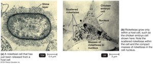
Rickettsia: Obligate intracellular parasites causing spotted fevers; transmitted by arthropods.
Ehrlichia: Tick-borne pathogens causing ehrlichiosis.
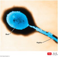
Caulobacter and Hyphomicrobium: Aquatic, reproduce by budding.
Rhizobium and Bradyrhizobium: Nitrogen-fixing symbionts of legumes.
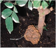
Agrobacterium: Plant pathogen causing crown gall disease.
Bartonella: Human pathogen (e.g., cat-scratch disease).
Brucella: Causes brucellosis, survives phagocytosis.
Nitrobacter and Nitrosomonas: Chemoautotrophs involved in nitrogen cycle.
Wolbachia: Endosymbionts affecting insect reproduction.
Betaproteobacteria
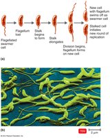
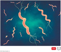
Spirillum: Freshwater, motile by polar flagella.
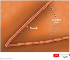
Sphaerotilus: Sheathed bacteria in freshwater and sewage.
Burkholderia: Degrades organic molecules; B. pseudomallei causes melioidosis.
Bordetella: Nonmotile rods; B. pertussis causes whooping cough.
Neisseria: Pathogens causing gonorrhea and meningitis.
Zoogloea: Important in wastewater treatment (activated sludge).
Gammaproteobacteria
Acidithiobacillus: Oxidizes sulfur compounds.
Thiotrichales (Beggiatoa): Aquatic, oxidizes sulfur for energy.
Francisella tularensis: Causes tularemia.
Pseudomonas: Opportunistic pathogens, metabolically diverse.
Azotobacter and Azomonas: Nitrogen-fixing bacteria.
Moraxella: Causes conjunctivitis.
Acinetobacter: Respiratory pathogen, antibiotic resistant.
Legionella: Causes legionellosis, found in water systems.
Coxiella burnetii: Causes Q fever, transmitted by aerosols or milk.
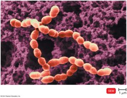
Vibrio: Aquatic, V. cholerae causes cholera.
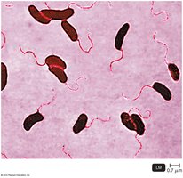
Enterobacteriales (Enterics): Facultative anaerobes, ferment carbohydrates, peritrichous flagella.
Escherichia coli: Indicator of fecal contamination, some strains pathogenic.
Salmonella: Foodborne illness, S. Typhi causes typhoid fever.
Shigella: Causes bacillary dysentery.
Klebsiella: Causes pneumonia.
Serratia: Red pigment, nosocomial infections.
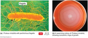
Proteus: Swarming motility, concentric ring colonies.
Yersinia pestis: Causes plague, transmitted by fleas.
Erwinia: Plant pathogens.
Enterobacter, Cronobacter: Nosocomial and foodborne pathogens.
Pasteurella: Animal pathogen, transmitted by bites.
Haemophilus: Requires X and V factors, causes meningitis and other infections.
Deltaproteobacteria
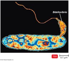
Bdellovibrio: Predatory on other Gram-negative bacteria.
Desulfovibrio: Sulfate-reducing, found in anaerobic environments.
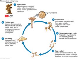
Myxococcales: Gliding motility, form fruiting bodies with myxospores.
Campylobacterota
Campylobacter: Microaerophilic, causes foodborne disease.
Helicobacter: Causes peptic ulcers and stomach cancer.
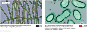
Cyanobacteria
Oxygenic photosynthetic bacteria, some fix nitrogen in heterocysts, important in Earth's oxygenation.
Chlorobi and Chloroflexi
Chlorobi: Green sulfur bacteria, anoxygenic photosynthesis.
Chloroflexi: Green nonsulfur bacteria.
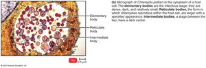
Chlamydiae
No peptidoglycan, obligate intracellular pathogens.
Chlamydia trachomatis: causes trachoma and urethritis.
Chlamydophila: causes respiratory infections.

Planctomycetes
Gram-negative, budding bacteria with unique cell wall and internal structures.
Gemmata obscuriglobus: has a membrane around DNA, resembling a eukaryotic nucleus.
Bacteroidota and Fusobacteria
Bacteroides: Anaerobic, abundant in human gut.
Cytophaga: Degrade cellulose and chitin.
Fusobacteria: Anaerobic, cause dental abscesses.
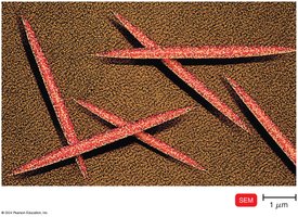
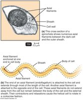
Spirochaetes
Coiled, motile via axial filaments.
Treponema pallidum: Causes syphilis.
Borrelia: Causes Lyme disease.
Leptospira: Excreted in animal urine.
Gram-Positive Bacteria
Divided by G+C content into Bacillota (low G+C) and Actinomycetota (high G+C).
Bacillota (Low G+C)
Clostridium: Endospore-forming, obligate anaerobes, cause tetanus, botulism, gas gangrene.
Clostridioides difficile: Causes antibiotic-associated diarrhea.
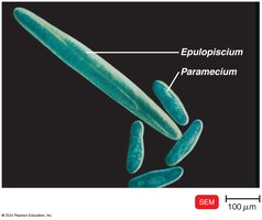
Epulopiscium: Large, daughter cells form within parent cell.
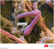
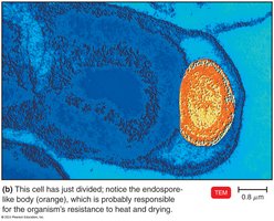
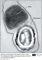
Bacillus: Endospore-forming rods, B. anthracis (anthrax), B. cereus (food poisoning).
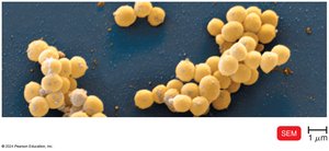
Staphylococcus: Grape-like clusters, S. aureus causes wound infections, often antibiotic resistant.
Lactobacillus: Used in food production, colonizes human body.
Streptococcus: Chains of cocci, cause pharyngitis, pneumonia, dental caries.
Enterococcus: Hospital contaminants, infect wounds and urinary tract.
Listeria monocytogenes: Foodborne pathogen.
Mycoplasma: Lack cell wall, pleomorphic, cause mild pneumonia.
Actinomycetota (High G+C)
Mycobacterium: Waxy cell wall, slow-growing, cause tuberculosis and leprosy.
Nocardia: Acid-fast, pulmonary infections.
Corynebacterium: C. diphtheriae causes diphtheria.
Propionibacterium: Forms propionic acid, C. acnes associated with acne.
Gardnerella vaginalis: Causes vaginitis.
Frankia: Nitrogen-fixing.
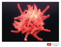
Streptomyces: Soil bacteria, produce antibiotics.
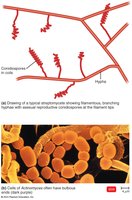
Actinomyces: Form filaments, found in mouth and throat.
Deinococcota
Deinococcus radiodurans: Highly radiation-resistant.
Thermus aquaticus: Source of Taq polymerase for PCR.
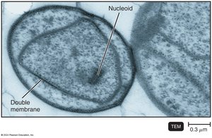
Archaea
Lack peptidoglycan, often extremophiles.
Halophiles: Require high salt.
Thermophiles: Grow at high temperatures.
Acidophiles: Grow at low pH.
Methanogens: Produce methane, some are human microbiota.
The Eukaryotes: Fungi, Algae, Protozoa, and Helminths
Fungi
Over 100,000 species; decomposers, pathogens, and beneficial organisms.
Form mycorrhizae with plants, produce antibiotics and food products.
Chemoheterotrophs, aerobic or facultative anaerobes.
Vegetative Structures
Hyphae: Filaments forming the mycelium.
Septate hyphae: With cross-walls.
Coenocytic hyphae: Without septa.
Yeasts: Unicellular, reproduce by budding or fission.
Dimorphic fungi: Yeastlike at 37°C, moldlike at 25°C.
Fungal Reproduction
Reproduce by sexual and asexual spores.
Asexual spores: Conidiospores, sporangiospores, etc.
Sexual spores: Formed by fusion of nuclei from two mating strains (plasmogamy, karyogamy, meiosis).
Medically Important Fungi
Mucoromycota: Conjugation fungi, e.g., Rhizopus.
Microsporidia: Obligate intracellular parasites, lack mitochondria.
Ascomycota: Sac fungi, produce ascospores.
Basidiomycota: Club fungi, produce basidiospores.
Fungal Diseases (Mycoses)
Systemic: Deep within body.
Subcutaneous: Beneath skin.
Cutaneous: Affect hair, skin, nails.
Superficial: Localized, e.g., hair shafts.
Opportunistic: Pathogenic in compromised hosts.
Algae
Photoautotrophs, mostly aquatic, lack true roots, stems, leaves.
Major groups: Brown, red, green algae, diatoms, dinoflagellates, euglenoids, oomycetes.
Important for oxygen production and as primary producers in aquatic ecosystems.
Protozoa
Unicellular eukaryotes, inhabit water and soil.
Complex life cycles, some are human pathogens (e.g., Plasmodium, Giardia).
Reproduce asexually (fission, budding, schizogony) and sexually (conjugation).
Some form cysts for survival.
Helminths
Multicellular parasitic worms: Platyhelminthes (flatworms) and Nematoda (roundworms).
Specialized for parasitism: reduced digestive, nervous, and locomotor systems; complex reproduction.
Life cycles may involve multiple hosts and larval stages.
Viruses, Viroids, and Prions
Distinctive Features of Viruses
Obligate intracellular parasites; require host cells to replicate.
Contain DNA or RNA, not both; protein coat (capsid), sometimes an envelope.
No ribosomes or ATP-generating mechanisms.
Viral Structure and Morphology
Virion: Complete viral particle.
Nucleic acid: DNA or RNA, single- or double-stranded, linear or circular.
Capsid: Protein coat made of capsomeres.
Envelope: Lipid, protein, and carbohydrate layer (in some viruses).
Spikes: Projections for attachment.
Viral Multiplication
Bacteriophages: Multiply via lytic or lysogenic cycles.
Lytic cycle: Attachment, penetration, biosynthesis, maturation, release.
Lysogenic cycle: Phage DNA integrates into host genome as a prophage.
Animal Virus Multiplication
Attachment, entry (endocytosis or fusion), uncoating, biosynthesis, maturation, release (budding or rupture).
Viral Genetics and Classification
Baltimore classification: Based on nucleic acid type and replication strategy.
Families end in -viridae, genera in -virus.
Viruses and Cancer
Some viruses are oncogenic, integrating into host DNA and causing transformation.
DNA oncogenic viruses: Adenoviridae, Herpesviridae, Papovaviridae, Hepadnaviridae.
RNA oncogenic viruses: Retroviridae (e.g., HTLV-1, HIV).
Viroids and Prions
Viroids: Short pieces of naked RNA, cause plant diseases.
Prions: Infectious proteins causing spongiform encephalopathies (e.g., mad cow disease, CJD).
Additional info: This guide covers the classification, structure, and diversity of prokaryotes (bacteria and archaea), eukaryotic microorganisms (fungi, algae, protozoa, helminths), and acellular infectious agents (viruses, viroids, prions), as outlined in standard microbiology curricula.
