 Back
BackThe Cell Cycle and Cell Division: Regulation, Mechanisms, and Implications
Study Guide - Smart Notes
 Tailored notes based on your materials, expanded with key definitions, examples, and context.
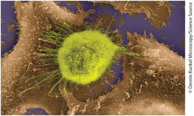
All Cells Derive from Other Cells
Overview of Cell Division
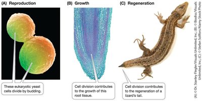
Cell division is a fundamental process in all living organisms, essential for reproduction, growth, and tissue repair. The process involves a series of coordinated events that ensure genetic material is accurately replicated and distributed to daughter cells.
Cell division signals: Initiate the process, responding to internal or external cues.
DNA replication: The genetic material is duplicated.
DNA segregation: Duplicated chromosomes are separated into daughter cells.
Cytokinesis: The cytoplasm divides, forming two distinct cells.
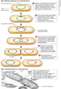
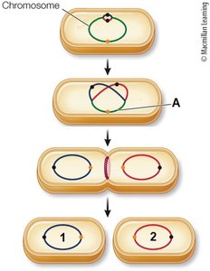
Prokaryotic Cell Division: Binary Fission
Prokaryotes, such as bacteria, reproduce by binary fission, a simple form of cell division resulting in two genetically identical cells.
Signals: Often external, such as nutrient availability.
Replication: Most prokaryotes have a single, circular chromosome. Replication begins at the ori (origin) and ends at the ter (terminus).
Segregation: The two ori regions move to opposite ends of the cell.
Cytokinesis: The cell membrane pinches in, and new cell wall material is synthesized, resulting in two separate cells.
Eukaryotic Cell Division
Eukaryotic cells divide in response to signals related to the organism's needs. The process is more complex due to multiple chromosomes and compartmentalization.
DNA replication: Occurs at multiple origins and is restricted to a specific cell cycle phase.
DNA segregation: Mitosis ensures each daughter cell receives one copy of each chromosome.
Cytokinesis: Differs between animal (membrane pinching) and plant cells (cell plate formation).
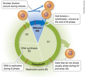
The Eukaryotic Cell Cycle Is Regulated
Phases of the Cell Cycle
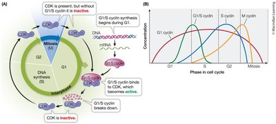
The eukaryotic cell cycle consists of interphase (G1, S, G2) and the M phase (mitosis and cytokinesis). Regulation ensures orderly progression and fidelity of division.
G1 phase: Cell grows and carries out normal functions; chromosomes are unreplicated.
S phase: DNA is replicated, producing sister chromatids.
G2 phase: Cell prepares for mitosis by synthesizing necessary components.
M phase: Includes mitosis (nuclear division) and cytokinesis (cytoplasmic division).
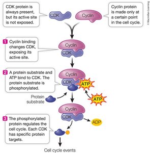
Regulation by Cyclin-Dependent Kinases (CDKs)
Progression through the cell cycle is controlled by cyclin-dependent kinases (CDKs), which are activated by binding to cyclins. This activation leads to phosphorylation of target proteins, driving cell cycle events.
CDKs: Protein kinases that require cyclin binding for activation.
Cyclins: Regulatory proteins whose levels fluctuate during the cell cycle.
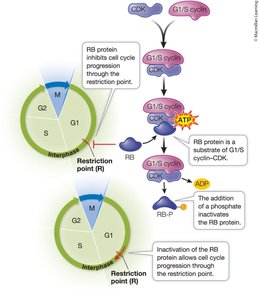
Checkpoints: Control points where the cell assesses readiness to proceed (e.g., restriction point in G1).
External Regulation and Growth Factors
External signals, such as growth factors, can stimulate cell division. These signals activate pathways that lead to cyclin synthesis and CDK activation.
Platelet-derived growth factor: Stimulates skin cell division for wound healing.
Interleukins and erythropoietin: Stimulate division and specialization of blood cells.
Eukaryotic Cells Divide by Mitosis
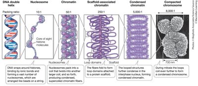
Chromosome Structure and Compaction
During mitosis, DNA is highly compacted into chromosomes, ensuring accurate segregation. Chromatin is organized by histones into nucleosomes, which further fold into higher-order structures.
Chromatin: DNA-protein complex forming chromosomes.
Nucleosomes: DNA wrapped around histone proteins.
Condensins: Proteins that help compact chromosomes during mitosis.
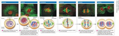
Phases of Mitosis
Mitosis is divided into distinct phases: prophase, prometaphase, metaphase, anaphase, and telophase. Each phase ensures the accurate distribution of chromosomes.
Prophase: Chromosomes condense, spindle apparatus forms.
Prometaphase: Nuclear envelope breaks down, spindle fibers attach to kinetochores.
Metaphase: Chromosomes align at the metaphase plate.
Anaphase: Sister chromatids separate and move to opposite poles.
Telophase: Nuclear envelopes reform around daughter chromosomes.
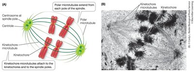
Spindle Apparatus and Chromosome Movement
The mitotic spindle, composed of microtubules, orchestrates chromosome movement. Centrosomes organize spindle poles, and kinetochores attach chromosomes to spindle fibers.
Centrosomes: Organize microtubules and define division plane.
Kinetochore microtubules: Attach to chromosomes and pull them apart.
Motor proteins: Kinesins and dyneins move chromosomes along microtubules.
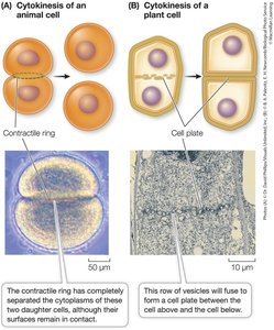
Cytokinesis
Cytokinesis divides the cytoplasm, completing cell division. The mechanism differs between animal and plant cells.
Animal cells: Contractile ring of actin and myosin pinches the cell in two.
Plant cells: Vesicles form a cell plate that develops into a new cell wall.
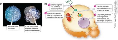
Cell Death Is Important in Living Organisms
Necrosis vs. Apoptosis
Cell death can occur by necrosis (uncontrolled) or apoptosis (programmed). Apoptosis is essential for development and tissue homeostasis.
Necrosis: Cell swells and bursts, causing inflammation.
Apoptosis: Cell contents are systematically dismantled and recycled without inflammation.

Unregulated Cell Division Can Lead to Cancer
Cancer and Tumor Formation
Cancer results from loss of cell cycle control, leading to continuous cell division and tumor formation. Tumors can be benign (localized) or malignant (invasive and metastatic).
Benign tumors: Grow slowly, resemble original tissue, and remain localized.
Malignant tumors: Invade surrounding tissues and can metastasize to distant sites.
Molecular Basis of Cancer
Cancer involves mutations in genes regulating the cell cycle. Oncogenes (overactive positive regulators) and tumor suppressors (inactive negative regulators) are key players.
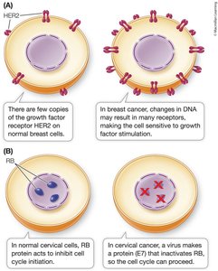
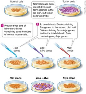
Oncogenes: Mutated genes that drive excessive cell division (e.g., HER2, Ras, Myc).
Tumor suppressors: Genes that inhibit cell division (e.g., RB, p53).
Multiple mutations: Often required for cancer development.
Cancer Treatments
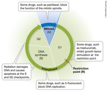
Treatments target rapidly dividing cells by interfering with the cell cycle. Strategies include surgery, chemotherapy, radiation, and targeted therapies.
Chemotherapy: Drugs like 5-fluorouracil and paclitaxel block DNA synthesis or spindle function.
Radiation: Damages DNA, inducing apoptosis in tumor cells.
Targeted therapy: Drugs like trastuzumab block specific growth factor receptors (e.g., HER2).
Summary Table: Comparison of Prokaryotic and Eukaryotic Cell Division
Feature | Prokaryotes | Eukaryotes |
|---|---|---|
Division Process | Binary fission | Mitosis (and meiosis in germ cells) |
Chromosome Structure | Single, circular | Multiple, linear |
Division Signals | External (e.g., nutrients) | Internal and external (developmental, growth factors) |
DNA Segregation | Simple, attached to membrane | Complex, involves spindle apparatus |
Cytokinesis | Membrane pinching, new wall formation | Contractile ring (animals), cell plate (plants) |
