Back
BackAmino Acids, Peptides, and Proteins: Structure, Properties, and Analysis
Study Guide - Smart Notes
 Tailored notes based on your materials, expanded with key definitions, examples, and context.
A. Structure and Properties of Amino Acids
1. General Structure and Hydrolysis of Proteins
Proteins, also known as polypeptides, polyamides, or peptides, are polymers composed of amino acid monomers. Complete hydrolysis of proteins yields their constituent amino acids. Most naturally occurring amino acids are α-amino acids, where the amino group is attached to the carbon atom adjacent (α-position) to the carboxyl group.
Protein hydrolysis breaks peptide bonds, releasing free amino acids.
α-Amino acids have the general structure: H2N–CHR–COOH, where R is the side chain.
2. Stereochemistry of Amino Acids
Most amino acids (except glycine) are chiral and exist in the L-configuration in nature. The L-form is determined by the position of the amino group on the left in a Fischer projection with the carboxyl group at the top.
Glycine is achiral (R = H).
L-amino acids are the biologically relevant isomers.
3. Classification of the 20 Proteinogenic Amino Acids
The 20 amino acids encoded by the genetic code are classified by the properties of their side chains:
Non-polar (hydrophobic): Found in protein interiors (e.g., Gly, Ala, Val, Leu, Ile, Phe, Trp, Met, Pro).
Polar (uncharged): Contain polar groups but are not ionized (e.g., Ser, Thr, Tyr, Cys, Asn, Gln).
Acidic: Negatively charged at physiological pH (e.g., Asp, Glu).
Basic: Positively charged at physiological pH (e.g., Lys, Arg, His).
B. Acid-Base Properties of Amino Acids
1. Ionization and pKa Values
Amino acids contain both an acidic carboxyl group and a basic amino group. Their ionization state depends on the pH of the solution:
At low pH: Both groups are protonated (cationic form).
At physiological pH: Carboxyl is deprotonated, amino is protonated (zwitterion).
At high pH: Both groups are deprotonated (anionic form).
The typical pKa values are:
Carboxyl group: ~2
Amino group: ~9–10
2. Isoelectric Point (pI)
The isoelectric point (pI) is the pH at which an amino acid has no net charge. For amino acids with neutral side chains, pI is the average of the carboxyl and amino group pKa values. For acidic or basic side chains, pI is the average of the two closest pKa values.
At pH > pI: Net negative charge.
At pH < pI: Net positive charge.
This property allows separation by electrophoresis:
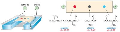
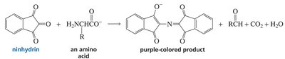
3. Detection of Amino Acids: Ninhydrin Reaction
Amino acids can be detected by reaction with ninhydrin, producing a purple color. This is used to visualize amino acids after separation techniques such as electrophoresis or chromatography.
C. Analytical Techniques for Amino Acids
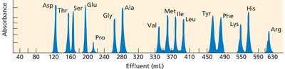
1. Ion-Exchange Chromatography
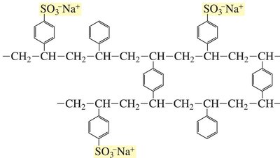
Ion-exchange chromatography separates amino acids based on their charge and hydrophobicity. The column is packed with a polymeric resin containing both negatively charged and hydrophobic groups. Positively charged amino acids adhere most strongly and elute last, while negatively charged and polar amino acids elute earlier.
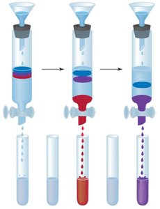
Fractions are collected and reacted with ninhydrin for quantification.
D. Protein Structure
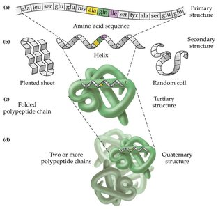
1. Primary Structure
The primary structure of a protein is the linear sequence of amino acids, written from the N-terminus (amino end) to the C-terminus (carboxyl end). The peptide bond has partial double-bond character due to resonance, restricting rotation and making the peptide group planar.
Sequence determines all higher levels of structure and function.
Peptide bonds are not basic and are planar.
2. Secondary Structure
Secondary structure refers to local folding patterns stabilized by hydrogen bonds between backbone atoms. The two most common motifs are the α-helix and the β-sheet.
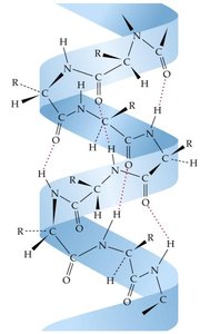
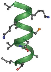
α-Helix
3.6 amino acids per turn.
Hydrogen bonds form between the NH group of one amino acid and the C=O group four residues earlier.
Right-handed helix; side chains project outward.

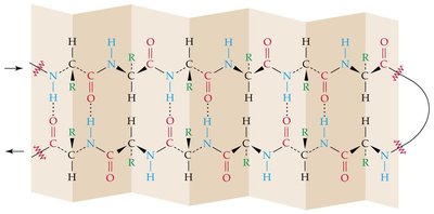
β-Sheet
Sheet-like arrangement of polypeptide chains.
Hydrogen bonds form between NH and C=O groups of adjacent chains.
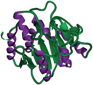
3. Tertiary Structure
Tertiary structure is the overall 3D folding of a single polypeptide chain, stabilized by hydrophobic interactions, hydrogen bonds, electrostatic interactions, and covalent disulfide bridges (formed by oxidation of cysteine thiols).
Loss of tertiary structure (denaturation) disrupts protein function.
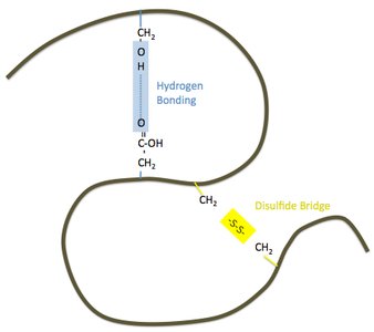
4. Hydrogen Bonds vs. Disulfide Bridges
Proteins such as keratin (in hair, nails, skin) derive their structure from both noncovalent hydrogen bonds and covalent disulfide bonds. Chemical treatments (e.g., hair curling) can alter disulfide bonds, affecting protein structure and properties.


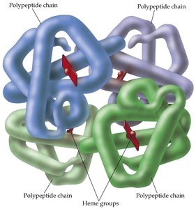
5. Quaternary Structure
Quaternary structure arises when two or more polypeptide subunits assemble into a functional protein complex. Hemoglobin is a classic example, consisting of four subunits. Proteins with only one polypeptide chain lack quaternary structure.