 Back
BackAmino Acids, Peptides, and Proteins: Structure, Properties, and Synthesis
Study Guide - Smart Notes
 Tailored notes based on your materials, expanded with key definitions, examples, and context.
Amino Acids and Peptides
Structure and Bonding
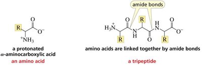
Amino acids are the fundamental building blocks of proteins and peptides. Each amino acid contains an amino group, a carboxylic acid group, a hydrogen atom, and a unique side chain (R group) attached to a central (α) carbon. Peptides and proteins are polymers of amino acids linked together by amide bonds (also called peptide bonds).
Amino acid: A molecule with both an amino group (–NH2) and a carboxylic acid group (–COOH) attached to the same carbon atom (the α-carbon).
Peptide bond: The amide linkage formed between the carboxyl group of one amino acid and the amino group of another.
Tripeptide: A peptide consisting of three amino acids joined by two peptide bonds.
Classification and Structures of Amino Acids
Common Amino Acids and Their Side Chains
The 20 standard amino acids differ only in their side chains (R groups), which determine their chemical properties and biological roles. They can be classified based on the nature of their side chains:
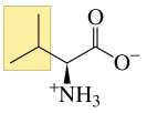
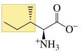
Aliphatic side chains: Nonpolar, hydrophobic (e.g., alanine, valine, leucine, isoleucine, glycine).
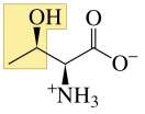
Hydroxy-containing: Polar, can form hydrogen bonds (e.g., serine, threonine).
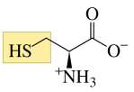
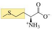
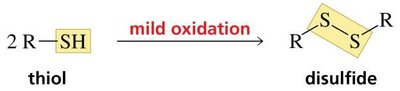
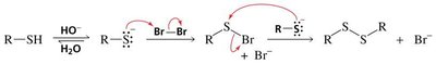
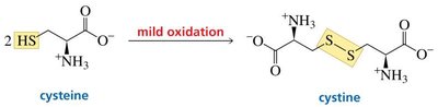
Sulfur-containing: Can form disulfide bonds (e.g., cysteine, methionine).
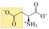
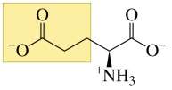
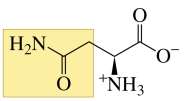
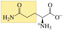
Acidic and amide derivatives: Negatively charged or polar (e.g., aspartate, glutamate, asparagine, glutamine).
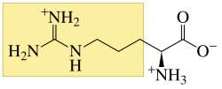
Basic: Positively charged at physiological pH (e.g., lysine, arginine, histidine).
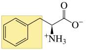
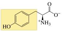
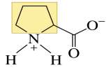
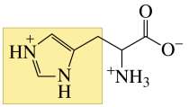
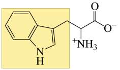
Aromatic and heterocyclic: Contain ring structures (e.g., phenylalanine, tyrosine, tryptophan, proline, histidine).

Special Features of Selected Amino Acids
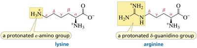
Lysine: Contains a protonated ε-amino group.
Arginine: Contains a protonated δ-guanidino group.
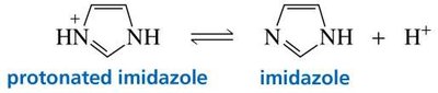
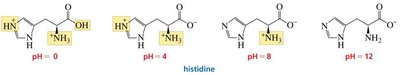
Histidine: Contains an imidazole ring, which can be protonated or neutral depending on pH.
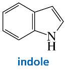
Tryptophan: Contains an indole ring.
Stereochemistry of Amino Acids
D- and L- Amino Acids
Amino acids (except glycine) are chiral and exist as D- and L- enantiomers. Proteins are composed exclusively of L-amino acids, which are related to L-glyceraldehyde by their configuration at the α-carbon.

Properties of Amino Acids
Acid-Base Properties and Zwitterions
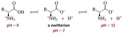
Amino acids can exist in different ionic forms depending on the pH of the solution. At physiological pH, they typically exist as zwitterions, with a positively charged amino group and a negatively charged carboxylate group.
At low pH: fully protonated (cationic form).
At neutral pH: zwitterion (no net charge).
At high pH: fully deprotonated (anionic form).
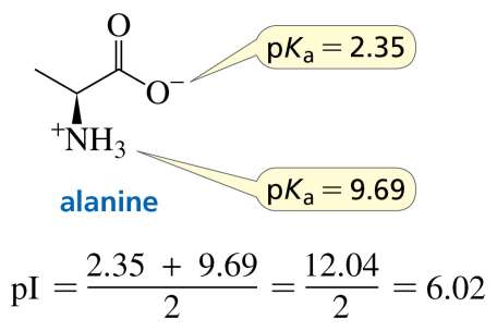
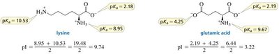
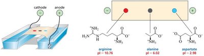
Isoelectric Point (pI)
The isoelectric point (pI) is the pH at which an amino acid has no net charge. For amino acids without ionizable side chains, the pI is the average of the pKa values of the carboxyl and amino groups:
Separation and Analysis of Amino Acids
Electrophoresis
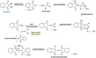
Electrophoresis separates amino acids based on their pI values. When placed in an electric field, amino acids migrate toward the electrode opposite their charge at a given pH. Ninhydrin is commonly used to detect amino acids after separation.
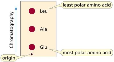
Chromatography
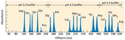
Chromatography separates amino acids based on their polarity. More polar amino acids travel less on the stationary phase, while less polar amino acids travel further.
Ion-Exchange Chromatography
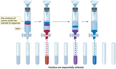
Ion-exchange chromatography uses a resin to separate amino acids based on their charge. Cation-exchange resins exchange sodium ions for positively charged amino acids, allowing for sequential elution and analysis.
Synthesis of Amino Acids
Laboratory Methods
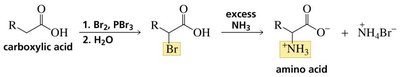
HVZ Reaction: Halogenation of a carboxylic acid followed by amination yields an α-amino acid.
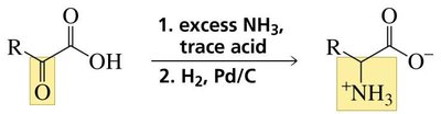
Reductive Amination: An α-keto acid is converted to an amino acid by reaction with ammonia and reduction.
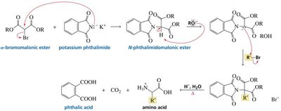
N-Phthalimidomalonic Ester Synthesis: Alkylation and hydrolysis of a phthalimidomalonic ester produces an amino acid.
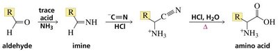
Strecker Synthesis: An aldehyde reacts with ammonia and cyanide, followed by hydrolysis, to yield an α-amino acid.
Peptides and Proteins: Structure and Synthesis
Peptide Bond Formation and Sequence
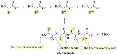
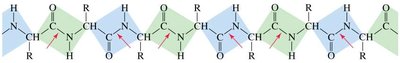
Peptides are formed by condensation reactions between amino acids, resulting in peptide bonds. The sequence of amino acids (primary structure) determines the properties and function of the peptide or protein.
N-terminal: The end of the peptide with a free amino group.
C-terminal: The end of the peptide with a free carboxyl group.
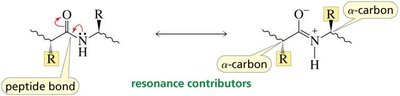
Peptide bonds have partial double-bond character due to resonance, restricting rotation.
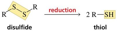
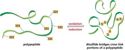
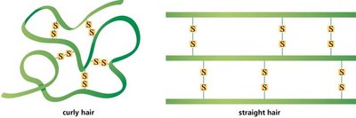
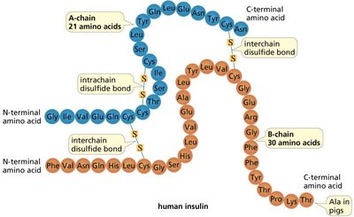
Disulfide Bonds and Protein Structure
Disulfide bonds (bridges) form between cysteine residues via oxidation, stabilizing protein structure. These covalent bonds are important for the tertiary and quaternary structure of proteins.
Summary Table: Properties of Selected Amino Acids
Name | Abbreviation | Side Chain Type | pKa (COOH) | pKa (NH3+) | pKa (Side Chain) |
|---|---|---|---|---|---|
Alanine | Ala, A | Aliphatic | 2.35 | 9.69 | N/A |
Arginine | Arg, R | Basic | 2.17 | 9.04 | 12.48 |
Aspartic acid | Asp, D | Acidic | 2.09 | 9.82 | 3.86 |
Cysteine | Cys, C | Sulfur-containing | 1.96 | 10.25 | 8.33 |
Glutamic acid | Glu, E | Acidic | 2.19 | 9.67 | 4.25 |
Histidine | His, H | Heterocyclic | 1.82 | 9.17 | 6.04 |
Lysine | Lys, K | Basic | 2.18 | 8.95 | 10.53 |
Tyrosine | Tyr, Y | Aromatic | 2.20 | 9.11 | 10.07 |
Key Concepts and Learning Objectives
Predict the charge of amino acids at different pH values.
Describe the isoelectric point, electrophoresis, and chromatography for amino acid analysis.
Draw peptides formed by reaction with exo- and endopeptidases.
Determine the amino acid sequence of a polypeptide.
Describe the primary, secondary, tertiary, and quaternary structures of proteins.
