 Back
BackNucleic Acids: Structure, Function, and Genetic Information Flow
Study Guide - Smart Notes
 Tailored notes based on your materials, expanded with key definitions, examples, and context.
Nucleic Acids
Primary Structure of Polynucleotides
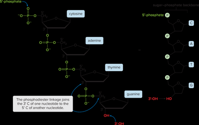
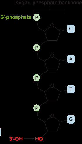
The primary structure of a polynucleotide refers to the linear sequence of nucleotides joined by phosphodiester bonds. Each nucleotide consists of a nitrogenous base (adenine, thymine, cytosine, or guanine in DNA), a deoxyribose sugar, and a phosphate group. The sequence is always read from the 5′ end to the 3′ end.
Phosphodiester Linkage: Connects the 3′-OH group of one nucleotide to the 5′-phosphate of the next.
Naming: The sequence CATG is read from the 5′ to 3′ direction.
Sugar-Phosphate Backbone: Provides structural stability to the nucleic acid strand.
Bases: Project from the backbone and encode genetic information.
Example: A DNA strand with the sequence 5′-CATG-3′.
The DNA Double Helix
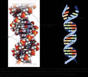
The double helix model of DNA, proposed by Watson and Crick with contributions from Rosalind Franklin, describes DNA as two antiparallel polynucleotide strands coiled into a right-handed helix. The sugar-phosphate backbones are on the outside, while the nitrogenous bases are on the inside, paired through hydrogen bonds.
Antiparallel Strands: One strand runs 5′ to 3′, the other 3′ to 5′.
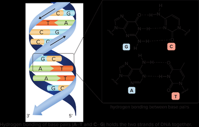
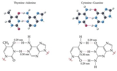
Base Pairing: Purines (adenine, guanine) pair with pyrimidines (thymine, cytosine).
Complementary Base Pairs: Adenine (A) pairs with Thymine (T) via two hydrogen bonds; Cytosine (C) pairs with Guanine (G) via three hydrogen bonds.
Stability: Hydrogen bonding and base stacking stabilize the helix.
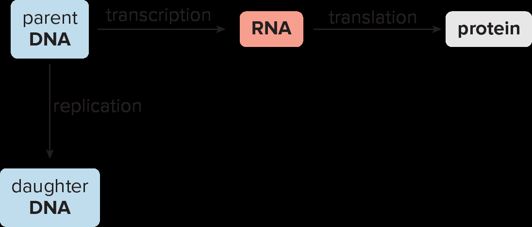
Flow of Genetic Information
Genetic information in DNA is used to direct protein synthesis through two key processes: transcription and translation. DNA replication ensures genetic continuity during cell division.
Replication: DNA makes a copy of itself before cell division.
Transcription: DNA is used as a template to synthesize RNA.
Translation: RNA directs the synthesis of proteins, determining the amino acid sequence.
DNA Replication
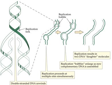
Mechanism of Replication
DNA replication is semiconservative: each new DNA molecule contains one parental and one newly synthesized strand. Replication begins at origins of replication, forming replication forks and bubbles.
Helicase: Unwinds the double helix at origins of replication.
Primase: Synthesizes short RNA primers to initiate DNA synthesis.
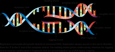
DNA Polymerase: Adds nucleotides to the 3′ end of the primer, synthesizing the new strand in the 5′ to 3′ direction.
Leading Strand: Synthesized continuously toward the replication fork.
Lagging Strand: Synthesized discontinuously as Okazaki fragments, later joined by DNA ligase.
Exonuclease: Removes RNA primers; DNA polymerase fills gaps.
DNA Ligase: Seals nicks in the sugar-phosphate backbone.
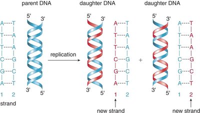
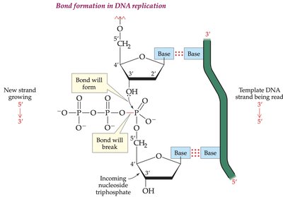
Base Pairing in DNA
During replication, the identity of the bases on the template strand determines the order of bases on the new strand. A pairs with T, and G pairs with C, ensuring accurate copying of genetic information.
RNA: Structure and Types
Differences Between DNA and RNA
RNA differs from DNA in several key aspects:
Sugar: RNA contains ribose; DNA contains deoxyribose.
Bases: RNA uses uracil (U) instead of thymine (T).
Structure: RNA is typically single-stranded and shorter than DNA.
Types: Three main types are ribosomal RNA (rRNA), messenger RNA (mRNA), and transfer RNA (tRNA).
Functions of RNA Types
rRNA: Forms the core of ribosomes, the site of protein synthesis.
mRNA: Carries genetic information from DNA to ribosomes.
tRNA: Brings specific amino acids to the ribosome during translation.
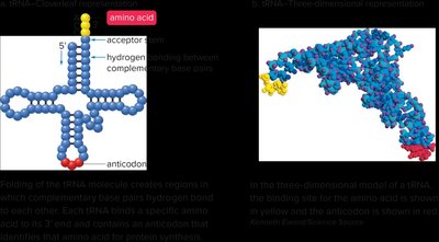
Structure of tRNA
tRNA molecules have a cloverleaf structure with an acceptor stem at the 3′ end (for amino acid attachment) and an anticodon loop (for codon recognition).
Transcription: DNA to RNA
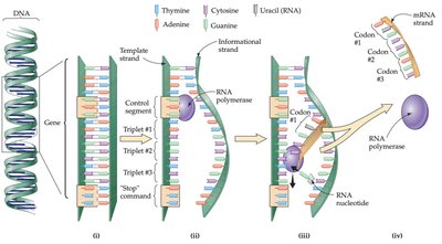
Mechanism of Transcription
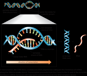
Transcription is the synthesis of mRNA from a DNA template. RNA polymerase binds to a promoter region, unwinds the DNA, and synthesizes mRNA complementary to the template strand.
Template Strand: Used to synthesize mRNA.
Informational (Coding) Strand: Has the same sequence as mRNA (except T is replaced by U).
Direction: mRNA is synthesized from the 5′ to 3′ direction.
Termination: Occurs when RNA polymerase reaches a stop signal.
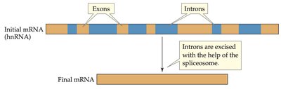
Processing of mRNA
In eukaryotes, the initial mRNA (heterogeneous nuclear RNA, hnRNA) contains both exons (coding regions) and introns (non-coding regions). Introns are removed by splicing before translation.
Exon: Codes for protein.
Intron: Non-coding, removed before translation.
Example: Transcription
Template strand: 3′—C T A G G A T A C—5′
mRNA: 5′—G A U C C U A U G—3′
Informational strand: 5′—G A T C C T A T G—3′
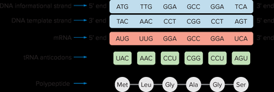
The Genetic Code
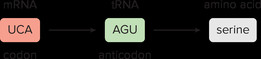
Codons and Amino Acids
The genetic code consists of triplets of nucleotides (codons) in mRNA, each specifying a particular amino acid. The code is universal and redundant (multiple codons can code for the same amino acid).
Start Codon: AUG (methionine) initiates translation.
Stop Codons: UAA, UAG, UGA signal termination of translation.
Example: UAC codes for tyrosine, UGC for cysteine.
mRNA Codon | tRNA Anticodon | Amino Acid |
|---|---|---|
ACA | UGU | Threonine |
GCG | CGC | Alanine |
AGA | UCU | Arginine |
UCC | AGG | Serine |
Translation and Protein Synthesis
Mechanism of Translation
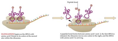
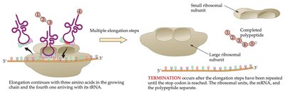
Translation is the process by which the sequence of codons in mRNA directs the synthesis of a polypeptide chain. It occurs in three main stages: initiation, elongation, and termination.
Initiation: mRNA binds to the ribosome; the first tRNA (carrying methionine) binds to the start codon (AUG).
Elongation: tRNAs bring amino acids to the ribosome, where peptide bonds form between them, extending the polypeptide chain.
Termination: When a stop codon is reached, the completed protein is released.

Mutations and Genetic Disease
Types of Mutations
A mutation is a change in the nucleotide sequence of DNA. Mutations can be spontaneous or induced by mutagens, and their effects vary depending on the type and location of the change.
Point Mutation: Substitution of one nucleotide for another.
Deletion Mutation: Loss of one or more nucleotides.
Insertion Mutation: Addition of one or more nucleotides.
Silent Mutation: No effect on the protein sequence due to redundancy in the genetic code.
Missense Mutation: Changes one amino acid in the protein; effect can be minor or severe (e.g., sickle cell anemia).
Genetic Disease: Mutations causing defective proteins that are inherited can lead to genetic diseases.
Disease | Characteristics |
|---|---|
Tay-Sachs disease | Mental retardation; defective hexosaminidase A enzyme |
Sickle cell anemia | Anemia; defective hemoglobin, capillary occlusion |
Phenylketonuria | Mental retardation; deficiency of phenylalanine hydroxylase |
Galactosemia | Mental retardation; deficiency of enzyme for galactose metabolism |
Huntington's disease | Progressive disability; defect in Htt protein gene, neuronal degeneration |
