 Back
Cell Division and Chromosome Heredity: Mitosis and Meiosis
Study Guide - Smart Notes
 Tailored notes based on your materials, expanded with key definitions, examples, and context.
Cell Division and Chromosome Heredity
Overview of Cell Division
Cell division is a fundamental process in genetics, responsible for the propagation of genetic material from one generation to the next. It occurs in two main forms: mitosis and meiosis. Mitosis produces genetically identical somatic cells, while meiosis generates gametes for sexual reproduction, each with half the chromosome number of the parent cell.
Somatic cells: Nonreproductive cells, typically diploid (2n).
Germ-line cells: Reproductive cells that undergo meiosis to produce gametes.
Diploid (2n): Two sets of chromosomes, one from each parent.
Haploid (n): One set of chromosomes, found in gametes.
Mitosis: Division of Somatic Cells
The Cell Cycle
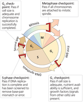
The cell cycle is a series of phases that regulate cell division and DNA replication. It consists of interphase (G1, S, G2) and the M phase (mitosis).
G1 phase: High gene expression and cell growth.
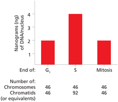
S phase: DNA replication, producing sister chromatids.
G2 phase: Preparation for cell division.
M phase: Cell division (mitosis or meiosis).
G0 phase: Cells exit the cycle, remain specialized, and do not divide.
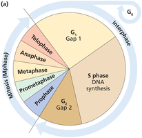
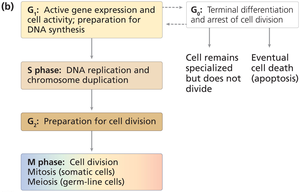
Substages of M Phase
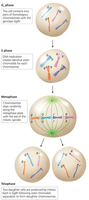
Mitosis is divided into five substages: prophase, prometaphase, metaphase, anaphase, and telophase. These stages ensure accurate partitioning of DNA (karyokinesis) and cytoplasmic contents (cytokinesis).
Prophase: Chromosome condensation begins.
Prometaphase: Nuclear envelope dissolves, spindle fibers attach to kinetochores.
Metaphase: Chromosomes align at the metaphase plate.
Anaphase: Sister chromatids separate and move to opposite poles.
Telophase: Nuclear membranes reform, chromosomes decondense.
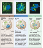
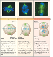
Chromosome Movement and Spindle Fibers
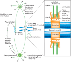
Centrosomes organize spindle fibers, which are essential for chromosome movement during mitosis. There are three types of spindle fibers:
Kinetochore microtubules: Attach to kinetochores and move chromosomes.
Polar microtubules: Extend toward the opposite pole, aiding cell elongation.
Astral microtubules: Grow toward the cell membrane, stabilizing the cell.
Sister Chromatid Cohesion and Separation
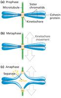
Cohesin proteins hold sister chromatids together, especially at the centromere. During anaphase, separase cleaves cohesin, allowing chromatids to separate.
Cohesin: Protein complex maintaining chromatid cohesion.
Separase: Enzyme that cleaves cohesin during anaphase.
Chromosome disjunction: Separation of sister chromatids.
Cytokinesis
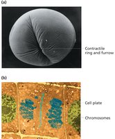
Cytokinesis divides the cytoplasm and organelles between daughter cells. In animal cells, a contractile ring forms a cleavage furrow; in plant cells, a cell plate forms.
Animal cells: Actin ring creates cleavage furrow.
Plant cells: Cell plate forms along the midline.
Genetic Consequences of Mitosis
Mitosis produces two genetically identical daughter cells, maintaining the diploid chromosome number. This ensures genetic continuity across generations.
Cell Cycle Checkpoints
Cell cycle checkpoints monitor readiness for progression. Mutations in checkpoint control can lead to abnormal cell growth, including cancer.
G1 checkpoint: Monitors cell size and DNA integrity.
S phase checkpoint: Ensures accurate DNA replication.
G2 checkpoint: Checks for cell size and DNA damage.
Metaphase checkpoint: Ensures chromosomes are properly attached to spindle fibers.
Meiosis: Production of Gametes for Sexual Reproduction
Overview of Meiosis
Meiosis is a specialized cell division process that produces four haploid gametes from a diploid cell. It consists of two sequential divisions: meiosis I and meiosis II. Meiosis introduces genetic variation through crossing over and independent assortment.
Asexual reproduction: Produces genetically identical offspring.
Sexual reproduction: Produces genetically diverse offspring via gametes.
Meiosis I: Homologous chromosomes separate, reducing chromosome number.
Meiosis II: Sister chromatids separate, producing four haploid cells.
Comparison of Mitosis and Meiosis
Feature | Mitosis | Meiosis |
|---|---|---|
Number of divisions | One | Two |
Number of daughter cells | Two | Four |
Chromosome number | Diploid (2n) | Haploid (n) |
Genetic identity | Identical | Unique |
Function | Growth, repair | Sexual reproduction |
Meiosis I: Hallmark Events
Meiosis I is characterized by homologous chromosome pairing, crossing over, and segregation. Prophase I is subdivided into leptotene, zygotene, pachytene, diplotene, and diakinesis.
Leptotene: Chromosome condensation begins.
Zygotene: Synapsis and formation of the synaptonemal complex.
Pachytene: Crossing over occurs at recombination nodules.
Diplotene: Chiasmata become visible as homologs pull apart.
Diakinesis: Chromosomes move toward the metaphase plate.
*Additional info: The synaptonemal complex is a protein structure essential for homolog pairing and recombination.*
Sex Chromosomes During Meiosis
X and Y chromosomes pair during male meiosis due to pseudoautosomal regions (PARs), allowing proper segregation.
Pseudoautosomal regions (PARs): Homologous regions on X and Y chromosomes.
Genetic Consequences of Meiosis
Meiosis generates genetic diversity through independent assortment and crossing over. The separation of homologs and chromatids forms the basis of Mendel's laws.
Law of Segregation: Each gamete receives one allele of each gene.
Law of Independent Assortment: Alleles of different genes assort independently.
Summary Table: Key Differences Between Mitosis and Meiosis
Process | Purpose | Chromosome Number | Genetic Variation |
|---|---|---|---|
Mitosis | Growth, repair | 2n → 2n | No |
Meiosis | Gamete production | 2n → n | Yes |
