 Back
BackCell Division and Chromosome Heredity: Mitosis and Meiosis
Study Guide - Smart Notes
 Tailored notes based on your materials, expanded with key definitions, examples, and context.
Cell Division and Chromosome Heredity
Introduction
Cell division is a fundamental process in genetics, ensuring the transmission of genetic material from one generation to the next. Two primary types of cell division are mitosis and meiosis, each serving distinct roles in growth, maintenance, and reproduction. Understanding these processes is essential for grasping the molecular basis of heredity and variation.
Mitosis: Division of Somatic Cells
Overview of Mitosis
Mitosis is the process by which a single somatic cell divides to produce two genetically identical daughter cells. This process is crucial for growth, tissue repair, and asexual reproduction in multicellular organisms. Mitosis maintains the diploid chromosome number (2n) in daughter cells.
Occurs in: Somatic (non-reproductive) cells
Product: Two genetically identical diploid cells
Genetic stability: Maintains chromosome number and genetic identity
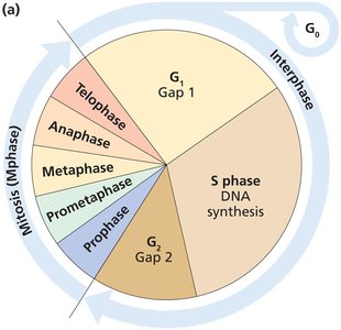
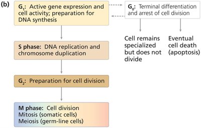
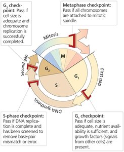
The Cell Cycle
The cell cycle is the ordered sequence of events that a cell undergoes from its formation to its division. It consists of interphase (G1, S, G2) and the M phase (mitosis and cytokinesis).
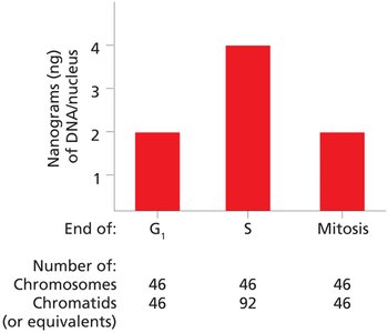
G1 phase: Cell growth and preparation for DNA synthesis
S phase: DNA replication and chromosome duplication
G2 phase: Preparation for cell division
M phase: Mitosis (nuclear division) and cytokinesis (cytoplasmic division)
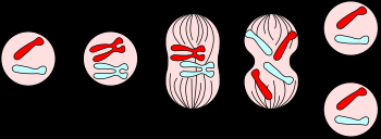
Substages of Mitosis (M Phase)
Mitosis is divided into five substages, each with distinct chromosomal and cellular events:
Prophase: Chromosomes condense, spindle apparatus forms
Prometaphase: Nuclear envelope breaks down, spindle fibers attach to kinetochores
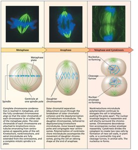
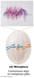
Metaphase: Chromosomes align at the metaphase plate
Anaphase: Sister chromatids separate and move to opposite poles
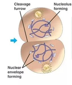
Telophase: Nuclear envelopes reform, chromosomes decondense
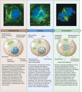
Chromosome Structure and Movement
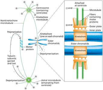
During mitosis, chromosomes undergo condensation and are manipulated by the mitotic spindle, composed of microtubules emanating from centrosomes. Key structures include:
Centrosome: Microtubule organizing center
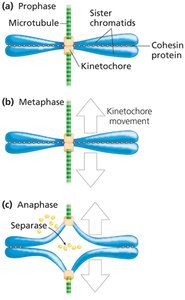
Centromere: Region where sister chromatids are joined
Kinetochore: Protein complex for microtubule attachment
Cohesin: Protein complex holding sister chromatids together
Sister Chromatid Cohesion and Separation
Cohesin proteins maintain sister chromatid cohesion until anaphase, when separase cleaves cohesin, allowing chromatids to separate.
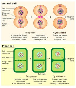
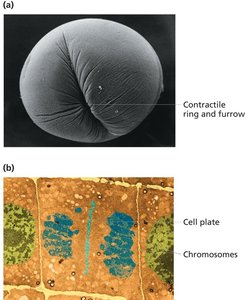
Completion of Mitosis and Cytokinesis
During telophase, nuclear envelopes reform and chromosomes decondense. Cytokinesis divides the cytoplasm, resulting in two identical daughter cells.
Genetic Consequences of Mitosis
Mitosis ensures genetic continuity by producing daughter cells with identical genetic material and chromosome number as the parent cell.
Cell Cycle Checkpoints
Checkpoints regulate the cell cycle, ensuring proper DNA replication and division:
G1 checkpoint: Monitors cell size, nutrients, and growth signals
S-phase checkpoint: Ensures complete and accurate DNA replication
G2 checkpoint: Verifies DNA replication and cell size
Metaphase checkpoint: Confirms chromosome attachment to spindle
Meiosis: Division for Sexual Reproduction
Overview of Meiosis
Meiosis is the process by which diploid germ-line cells divide to produce haploid gametes (sperm and eggs). It consists of two sequential divisions (meiosis I and II) following a single round of DNA replication, resulting in four genetically unique haploid cells.
Occurs in: Germ-line cells
Product: Four genetically distinct haploid cells
Genetic diversity: Generated by crossing over and independent assortment
Comparison of Mitosis and Meiosis
Characteristic | Mitosis | Meiosis |
|---|---|---|
Purpose | Growth, maintenance | Gamete production, genetic diversity |
Location | Somatic cells | Germ-line cells |
Divisions | One | Two (I & II) |
Product | 2 identical diploid cells | 4 unique haploid cells |
Homologous chromosomes | No pairing | Pairing, crossing over, separation |
Sister chromatids | Separate in anaphase | Separate in meiosis II |
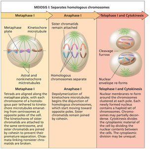
Meiosis I: Reductional Division
Meiosis I separates homologous chromosomes, reducing the chromosome number by half. Key events include:
Prophase I: Homologous chromosomes pair (synapsis) and exchange genetic material (crossing over)
Metaphase I: Homologous pairs align at the metaphase plate
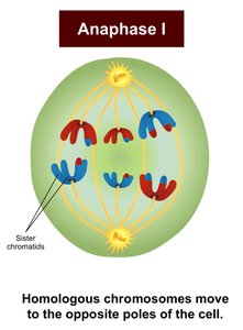
Anaphase I: Homologs separate to opposite poles
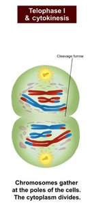
Telophase I and Cytokinesis: Two haploid cells form
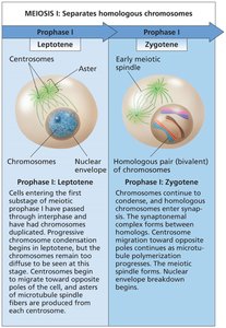
Prophase I Substages
Leptotene: Chromosome condensation begins
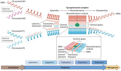
Zygotene: Synapsis and formation of synaptonemal complex
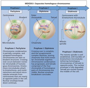
Pachytene: Crossing over occurs
Diplotene: Homologs begin to separate, chiasmata visible
Diakinesis: Chromosomes prepare for metaphase I
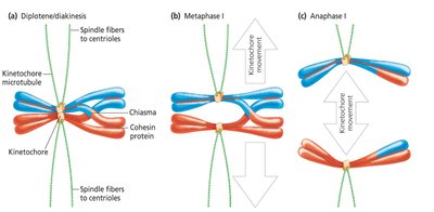
Metaphase I and Anaphase I
By metaphase I, homologs are aligned and chiasmata are resolved. In anaphase I, homologous chromosomes (not sister chromatids) are separated.
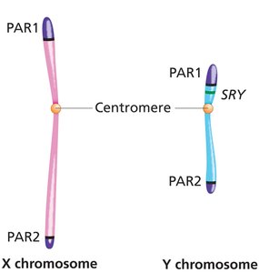
Sex Chromosomes During Meiosis
Despite limited homology, X and Y chromosomes pair during male meiosis due to pseudoautosomal regions (PARs) present on both chromosomes, allowing synapsis and recombination.
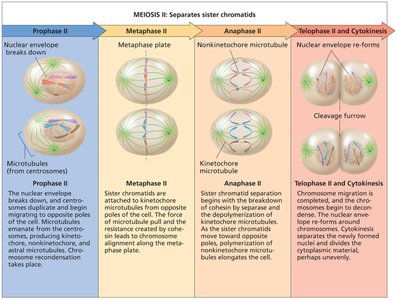
Meiosis II: Equational Division
Meiosis II resembles mitosis, separating sister chromatids in each haploid cell to produce four genetically distinct haploid gametes.
Genetic Consequences of Meiosis
Meiosis generates genetic diversity through independent assortment and crossing over, providing the mechanical basis for Mendel’s laws of segregation and independent assortment. For example, in a heterozygote (Aa), homologs bearing A and a alleles segregate during anaphase I, resulting in gametes with a 1:1 ratio of alleles.
Key Terms: Mitosis, meiosis, chromosome, chromatid, centromere, kinetochore, cohesin, synapsis, crossing over, chiasma, pseudoautosomal region, gamete, zygote, diploid, haploid.
