 Back
BackCell Division and Chromosome Heredity: Mitosis, Meiosis, and Chromosomal Basis of Heredity
Study Guide - Smart Notes
 Tailored notes based on your materials, expanded with key definitions, examples, and context.
Cell Division and Chromosome Heredity
Overview of Cell Division
Cell division is a fundamental process in genetics, enabling growth, maintenance, and reproduction in organisms. Two main types of cell division are mitosis and meiosis, each serving distinct biological purposes and producing different cellular outcomes.
Mitosis: Produces two genetically identical daughter cells for growth and tissue maintenance.
Meiosis: Produces four genetically distinct haploid cells for sexual reproduction.
The Cell Cycle
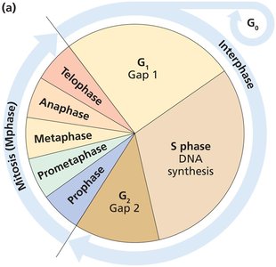
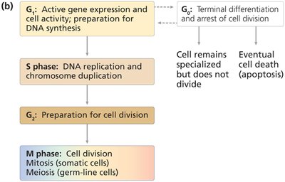
The cell cycle is the series of events that cells undergo as they grow and divide. It consists of interphase (G1, S, G2) and the M phase (mitosis or meiosis).
G1 phase: Cell growth and preparation for DNA synthesis.
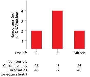
S phase: DNA replication and chromosome duplication.
G2 phase: Preparation for cell division.
M phase: Cell division (mitosis or meiosis).
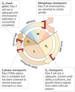
Major Cell Cycle Checkpoints
Checkpoints ensure the fidelity of cell division by monitoring DNA integrity, chromosome attachment, and cell size.
G1 checkpoint: Monitors cell size, nutrients, and growth signals.
S-phase checkpoint: Ensures DNA replication is complete and error-free.
G2 checkpoint: Checks for adequate cell size and successful chromosome replication.
Metaphase checkpoint: Ensures all chromosomes are attached to the mitotic spindle.
Mitosis: Division of Somatic Cells
Stages of Mitosis
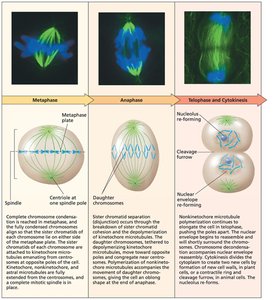
Mitosis is divided into five substages: prophase, prometaphase, metaphase, anaphase, and telophase. It results in the partitioning of DNA and cytoplasmic contents into two daughter cells.
Prophase: Chromosome condensation begins.
Prometaphase: Nuclear envelope breaks down; spindle fibers attach to kinetochores.
Metaphase: Chromosomes align at the metaphase plate.
Anaphase: Sister chromatids separate and move to opposite poles.
Telophase: Nuclear membranes reassemble; chromosomes decondense.
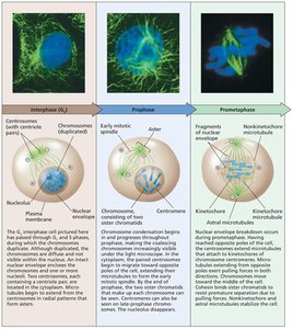
Chromosome Condensation and Structure
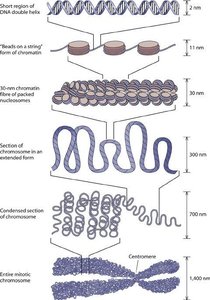
Chromosomes undergo progressive condensation during mitosis, reaching maximum compaction at metaphase. Chromatin structure is organized at multiple levels, from DNA double helix to fully condensed mitotic chromosomes.
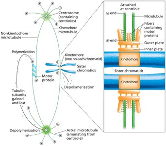
Microtubules and Chromosome Movement
Microtubules emanate from centrosomes and are classified as kinetochore, polar, and astral microtubules. Kinetochore microtubules attach to chromosomes and facilitate their movement during mitosis.
Kinetochore microtubules: Attach to kinetochores on chromosomes.
Polar microtubules: Interact with microtubules from the opposite pole.
Astral microtubules: Anchor the spindle apparatus to the cell membrane.
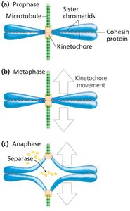
Sister Chromatid Cohesion and Separation
Cohesin proteins maintain sister chromatid cohesion until anaphase, when separase cleaves cohesin, allowing chromatids to separate.
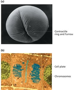
Cytokinesis
Cytokinesis is the final step of cell division, partitioning the cytoplasm into two daughter cells. In animal cells, a contractile ring forms a cleavage furrow; in plant cells, a cell plate forms.
Meiosis: Division for Sexual Reproduction
Overview and Comparison to Mitosis
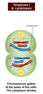
Meiosis consists of two sequential divisions (meiosis I and II) following a single round of DNA replication. It produces four genetically distinct haploid cells, providing the basis for genetic diversity in sexual reproduction.
Meiosis I: Homologous chromosomes pair, undergo crossing over, and segregate.
Meiosis II: Sister chromatids separate, similar to mitosis.
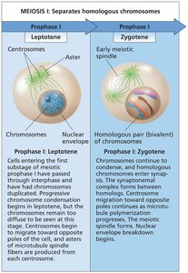
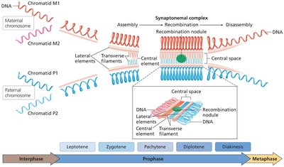
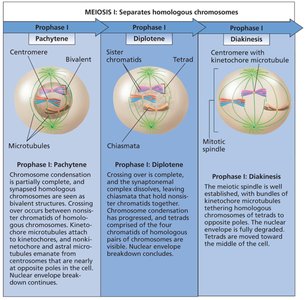
Meiosis I: Homologous Chromosome Pairing and Recombination
Meiosis I is characterized by homologous chromosome pairing, synapsis, and crossing over, which increases genetic variation.
Leptotene: Chromosome condensation begins.
Zygotene: Synapsis and formation of the synaptonemal complex.
Pachytene: Crossing over occurs between nonsister chromatids.
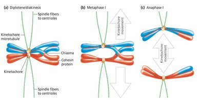
Diplotene: Chiasmata become visible; homologs begin to separate.
Diakinesis: Chromosomes fully condense; spindle forms.
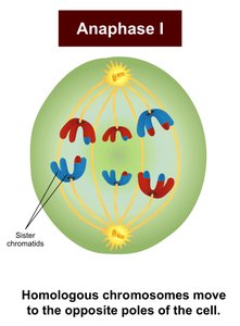
Homolog Separation and Chromosome Movement
During metaphase I, homologs align at the metaphase plate, and chiasmata are resolved. In anaphase I, homologous chromosomes move to opposite poles, reducing chromosome number by half.
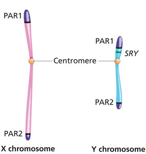
Sex Chromosomes and Pseudoautosomal Regions
X and Y chromosomes pair during meiosis due to pseudoautosomal regions (PARs), which allow synapsis and recombination.
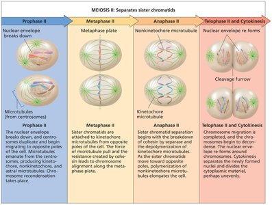
Meiosis II: Separation of Sister Chromatids
Meiosis II resembles mitosis, separating sister chromatids to produce four haploid cells.
Genetic Basis of Mendel's Laws
Meiosis provides the mechanical basis for Mendel's laws of segregation and independent assortment. Homologs bearing different alleles separate during anaphase I, generating Mendelian ratios in gametes.
Chromosomal Basis of Heredity
The Chromosome Theory of Heredity
The chromosome theory, proposed by Sutton and Boveri, states that genes are carried on chromosomes, and their behavior during meiosis mirrors hereditary transmission.
Wild type: The most common phenotype in a population.
Sex-linked inheritance: Transmission of genes on sex chromosomes.
Sex Determination Mechanisms
Sex determination can be chromosomal or genetic, depending on the species. In mammals, the presence of the SRY gene on the Y chromosome initiates male development.
Chromosomal sex: Determined at fertilization by the presence of X and Y chromosomes.
Phenotypic sex: Determined by gene expression and morphology.
X/A ratio: In Drosophila, the ratio of X chromosomes to autosome sets determines sex (male: 0.5, female: 1.0).
Diversity of Sex Determination Systems
Other organisms use different systems, such as the Z/W system in birds, reptiles, and some fish, where females are ZW and males are ZZ.
Human Sex-Linked Transmission
X-Linked Recessive and Dominant Inheritance
X-linked traits display distinct inheritance patterns. Recessive traits are more frequently expressed in males due to hemizygosity, while dominant traits are transmitted differently between sexes.
X-linked recessive: Trait expressed more often in males; can skip generations.
X-linked dominant: Heterozygous females transmit the trait to half their offspring; dominant males transmit to all daughters.
Y-linked: Exclusively male-to-male transmission.
Dosage Compensation
Mechanisms of Dosage Compensation
Dosage compensation equalizes the expression of sex-linked genes between males and females. Different organisms use distinct mechanisms:
Animal | Sex Chromosomes (Males) | Sex Chromosomes (Females) | Dosage Compensation Mechanism |
|---|---|---|---|
Fruit fly | X Y | X X | Expression of X-linked genes in males is doubled relative to females. |
Roundworm | X O | X X | Gene expression of each X chromosome in hermaphrodites is decreased to half that of males. |
Marsupial mammals | X Y | X X | Paternally derived X chromosome is inactivated in all female somatic cells. |
Placental mammals | X Y | X X | One X chromosome is randomly inactivated in each female somatic cell. |
X-Chromosome Inactivation in Mammals
In placental mammals, one X chromosome in each female somatic cell is randomly inactivated, forming a Barr body. This results in females being mosaics of cells expressing either the maternal or paternal X chromosome.
Mechanism of X Inactivation
X inactivation is mediated by the XIST gene, which produces RNA that coats the chromosome to be inactivated. Some genes escape inactivation, and XIST acts only in cis.
Visible Mosaicism in Cats
Calico and tortoiseshell cats are examples of visible mosaicism due to X inactivation, resulting in unique patterns of orange and black patches.
