 Back
BackCell Division and Chromosome Heredity: Structure, Function, and Mitosis
Study Guide - Smart Notes
 Tailored notes based on your materials, expanded with key definitions, examples, and context.
Introduction to Chromosomes and Cell Division
Overview of Chromosomes
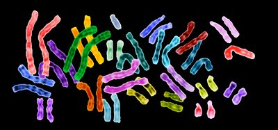
Chromosomes are essential structures within living cells that contain genetic material, primarily DNA, and associated proteins. Their organization and behavior during cell division are fundamental to understanding heredity and variation in genetics.
Chromosomes are composed of DNA (genetic material) and proteins (structural support).
In eukaryotes, the DNA-protein complex is called chromatin.
Chromosomes contain genes, the units of heredity.
Types of Cells: Prokaryotes vs. Eukaryotes
Cells are classified as prokaryotic or eukaryotic based on their internal structure and chromosome organization.
Prokaryotes (Bacteria, Archaea): No nucleus, usually a single circular chromosome in the nucleoid region.
Eukaryotes (Protists, fungi, plants, animals): Nucleus containing linear chromosomes, membrane-bound organelles (e.g., mitochondria, chloroplasts) with their own DNA.
General Features and Morphology of Chromosomes
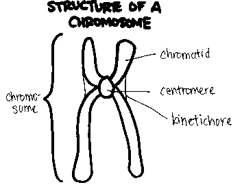
Chromosome Structure
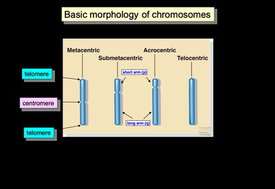
Chromosomes have distinct morphological features that are important for their identification and function during cell division.
Telomeres: Protective ends of chromosomes.
Centromere: Central region where sister chromatids are joined and spindle fibers attach during division.
Chromosomes are classified by centromere position: metacentric, submetacentric, acrocentric, and telocentric.
Chromosome Number and Species Variation
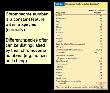
The number of chromosomes is a constant feature within a species and can be used to distinguish between species.
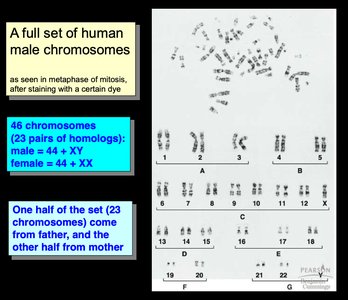
Humans: 46 chromosomes (23 pairs)
Other species have different chromosome numbers (e.g., dogs: 78, fruit flies: 8).
Organism | Total Chromosome Number |
|---|---|
Human | 46 |
Chimpanzee | 48 |
Dog | 78 |
Cat | 38 |
Mouse | 40 |
Chicken | 78 |
Fruit fly | 8 |
Tobacco | 48 |
Yeast | 32 |
Other species | Varies |
Genetic Similarity and Homologous Chromosomes
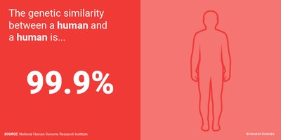
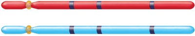
Members of a chromosome pair are called homologs. Homologous chromosomes are nearly identical in size, banding pattern, and gene content, but may carry different alleles.
Genetic similarity between humans is about 99.9%.
Allelic differences account for variation in traits (e.g., eye color).
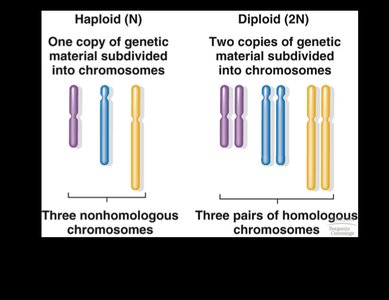
Diploid and Haploid Chromosome Sets
Eukaryotic organisms can be diploid (2N) or haploid (N), referring to the number of chromosome sets in their cells.
Diploid (2N): Two sets of chromosomes (e.g., humans: 46 chromosomes, 23 pairs).
Haploid (N): One set of chromosomes (e.g., gametes: sperm and egg).
Homologous Chromosomes and Gene Loci
The physical location of a gene on a chromosome is called its locus. Homologous chromosomes carry the same genes at the same loci, but may have different alleles.
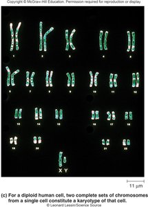
Karyotype Analysis
A karyotype is an organized profile of a species' chromosomes, used to detect abnormalities and distinguish between species.
Human karyotype: 46 chromosomes, including sex chromosomes (XX or XY).
One set of chromosomes is inherited from each parent.
Cell Division: Mitosis and the Cell Cycle
Purpose and Significance of Cell Division
Cell division is essential for reproduction, growth, development, and maintenance of multicellular organisms. It occurs via mitosis in somatic cells and meiosis in germ cells.
Asexual reproduction: Unicellular organisms produce new individuals by cell division (e.g., bacteria, yeast).
Multicellularity: Development from a single fertilized egg to a complex organism.
Mitosis: Enables growth, tissue repair, and replacement of cells.
Binary fission: Prokaryotic cell division.
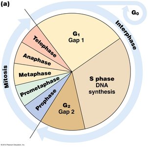
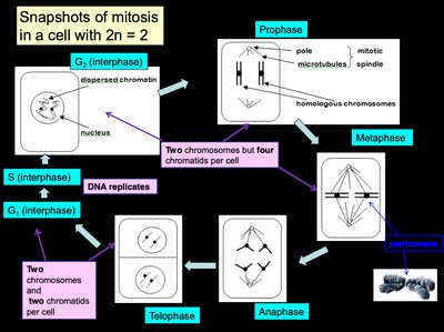
The Cell Cycle
The cell cycle is a regulated sequence of events that includes DNA replication and cell division. It consists of interphase (G1, S, G2) and the M phase (mitosis and cytokinesis).
G1 phase: Cell growth and gene expression.
G0 phase: Non-dividing, specialized state.
S phase: DNA synthesis and replication.
G2 phase: Preparation for division.
M phase: Mitosis (nuclear division) and cytokinesis (cytoplasmic division).

Mitosis: Stages and Chromosome Behavior
Substages of Mitosis
Mitosis is a dynamic process divided into several substages, each characterized by specific chromosomal and cellular events.
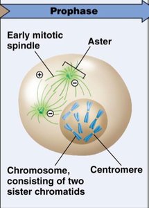
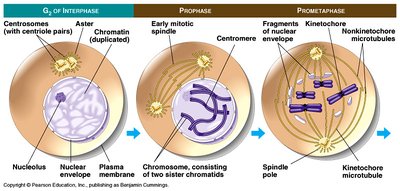
Prophase: Chromosome condensation, disappearance of nucleolus, centrosome migration, spindle formation.
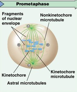
Prometaphase: Nuclear envelope breakdown, spindle fibers attach to kinetochores, chromosomes move to cell center.
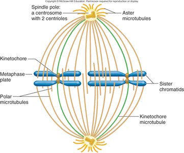
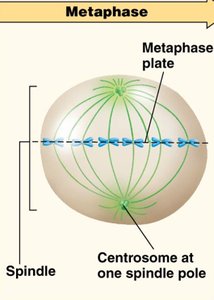
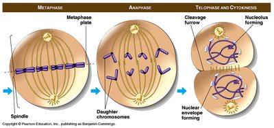
Metaphase: Chromosomes align at the metaphase plate, maximum condensation.
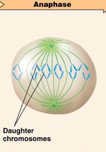
Anaphase: Sister chromatids separate and move to opposite poles.
Telophase: Chromosomes decondense, nuclear envelopes reform.
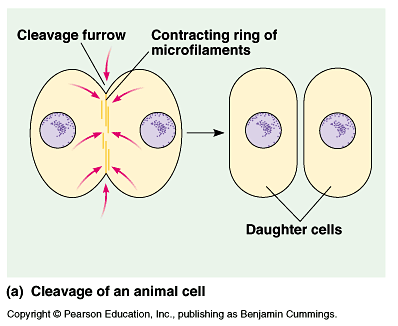
Cytokinesis: Division of cytoplasm, formation of two daughter cells.
Chromosome Condensation and Distribution
During mitosis, chromosomes condense and become visible. Centrosomes organize spindle fibers that facilitate chromosome movement and segregation.
Centromeres: Specialized DNA sequences joining sister chromatids.
Kinetochores: Protein complexes at centromeres for spindle attachment.
Spindle fibers: Microtubules responsible for chromosome movement.
Detailed Stages of Mitosis
Prophase
Chromosomes condense, nucleolus disappears, spindle apparatus begins to form.
Prometaphase
Nuclear envelope fragments, spindle fibers attach to kinetochores, chromosomes begin to move.
Metaphase
Chromosomes align at the metaphase plate, spindle fibers fully attached.
Anaphase
Sister chromatids separate and are pulled to opposite poles, ensuring equal genetic material in daughter cells.
Telophase and Cytokinesis
Chromosomes decondense, nuclear envelopes reform, and the cytoplasm divides to produce two genetically identical daughter cells.
Example: Mitosis in Plant Cells
Mitosis in plant cells follows the same stages as in animal cells, but cytokinesis occurs via cell plate formation rather than cleavage.
Summary Table: Key Differences in Cell Division
Feature | Prokaryotes | Eukaryotes |
|---|---|---|
Chromosome Type | Circular | Linear |
Division Process | Binary Fission | Mitosis/Meiosis |
Nucleus | Absent | Present |
Organelles | Absent | Present |
Key Terms and Concepts
Chromosome: Structure containing DNA and proteins, carrying genetic information.
Homologous chromosomes: Chromosome pairs with similar structure and gene content.
Karyotype: Complete set of chromosomes in a cell.
Mitosis: Process of nuclear division producing genetically identical cells.
Cytokinesis: Division of cytoplasm following mitosis.
Centromere: Region joining sister chromatids.
Kinetochores: Protein complexes for spindle attachment.
Spindle fibers: Microtubules facilitating chromosome movement.
Equations and Formulas
Chromosome Number in Diploid Cells:
DNA Replication:
Additional info: Academic context and definitions have been expanded for clarity and completeness. All images included are directly relevant to the adjacent explanations and reinforce key concepts in chromosome structure, cell division, and mitosis.
