 Back
BackCell Division and Chromosome Heredity: Structured Study Notes
Study Guide - Smart Notes
 Tailored notes based on your materials, expanded with key definitions, examples, and context.
Cell Division and Chromosome Heredity
Overview of Cell Division
Cell division is a fundamental process in all living organisms, enabling growth, development, tissue renewal, and reproduction. Eukaryotic cell division occurs via two main mechanisms: mitosis and meiosis. Mitosis produces genetically identical somatic cells, while meiosis generates genetically diverse gametes for sexual reproduction.
Mitosis: Division of the nucleus and cytoplasm in somatic cells.
Meiosis: Specialized division in germ-line cells, resulting in haploid gametes.
Key Roles of Cell Division
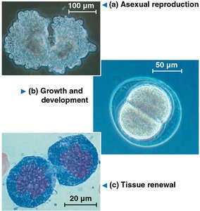
Cell division serves several essential functions in multicellular organisms:
Asexual reproduction: Produces offspring without genetic variation.
Growth and development: Increases cell number during organismal development.
Tissue renewal: Replaces damaged or dead cells.
Types of Cell Division
Mitosis
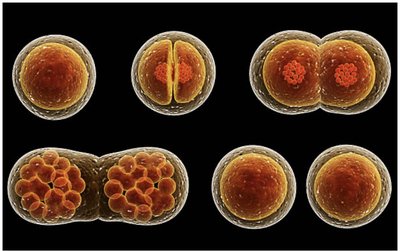
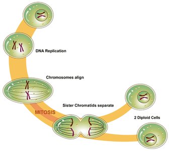
Mitosis is the process by which somatic cells divide, resulting in two genetically identical daughter cells. It involves a single round of DNA replication followed by one division.
Purpose: Growth, maintenance, and repair.
Product: Two diploid cells.
Meiosis
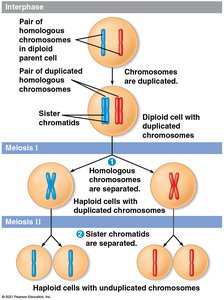
Meiosis is a two-stage division process in germ-line cells, producing four genetically distinct haploid gametes. It is essential for sexual reproduction and genetic diversity.
Purpose: Formation of gametes (sperm and egg).
Product: Four haploid cells.
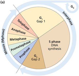
The Cell Cycle
Structure and Progression
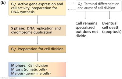
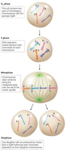
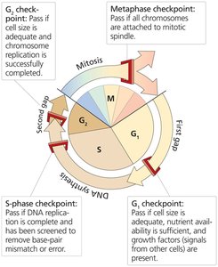
The cell cycle consists of interphase (G1, S, G2), G0 phase, and M phase. Interphase is the period of cell growth and DNA replication, while M phase includes mitosis and cytokinesis.
G1 phase: Cell growth and preparation for DNA synthesis.
S phase: DNA replication and chromosome duplication.
G2 phase: Preparation for cell division.
G0 phase: Terminal differentiation; cells exit the cycle.
M phase: Cell division (mitosis or meiosis).
Mitosis: Division of Somatic Cells
Stages of Mitosis
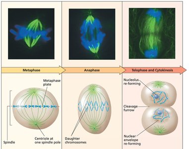
Mitosis is divided into five stages: prophase, prometaphase, metaphase, anaphase, and telophase. Each stage is characterized by specific events involving chromosomes and spindle fibers.
Prophase: Chromosomes condense, spindle forms.
Prometaphase: Nuclear envelope breaks down, spindle fibers attach to kinetochores.
Metaphase: Chromosomes align at the metaphase plate.
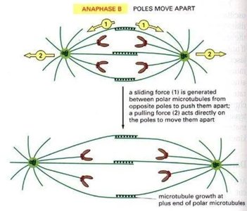
Anaphase: Sister chromatids separate and move to opposite poles.
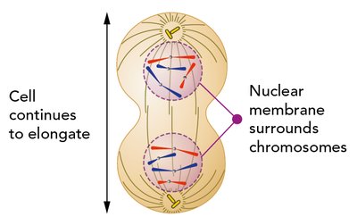
Telophase: Nuclear envelope reforms, chromosomes decondense.
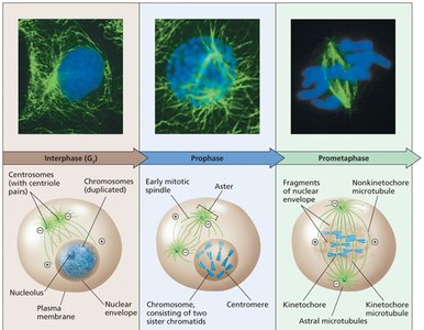
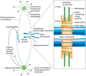
Types of Spindle Fibers
Spindle fibers are essential for chromosome movement and cell stability during mitosis:
Kinetochore microtubules: Attach to kinetochores at centromeres, move chromosomes.
Polar microtubules: Extend toward opposite poles, elongate cell.
Astral microtubules: Grow toward cell membrane, stabilize cell.
Sister Chromatid Cohesion
Sister chromatids are held together by cohesin proteins until anaphase, when separase cleaves cohesin, allowing chromatids to separate.
Cohesin: Protein complex maintaining chromatid cohesion.
Separase: Enzyme that cleaves cohesin during anaphase.
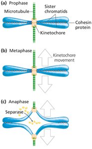
Anaphase and Completion of Cell Division
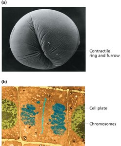
During anaphase, spindle fibers pull sister chromatids apart. Telophase and cytokinesis complete cell division, resulting in two identical daughter cells.
Anaphase: Chromatids move to opposite poles.
Telophase: Nuclear envelope reforms.
Cytokinesis: Cytoplasm divides.
Mitosis Produces Identical Daughter Cells
Mitosis ensures genetic consistency by producing two daughter cells with identical DNA content.
Genetic identity: Maintained through accurate chromosome segregation.
Cell Cycle Checkpoints
Checkpoints regulate the cell cycle, ensuring proper DNA replication and chromosome segregation.
G1 checkpoint: Monitors cell size and nutrient availability.
S-phase checkpoint: Ensures complete and accurate DNA replication.
G2 checkpoint: Verifies DNA integrity before mitosis.
Metaphase checkpoint: Confirms chromosome attachment to spindle.
Meiosis: Production of Gametes
Stages of Meiosis I and II
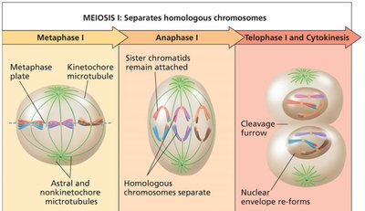
Meiosis consists of two sequential divisions: Meiosis I (reductional) and Meiosis II (equational). Key events in Meiosis I include homologous chromosome pairing, crossing over, and segregation.
Meiosis I: Homologous chromosomes pair, undergo crossing over, and segregate.
Meiosis II: Sister chromatids separate, similar to mitosis.
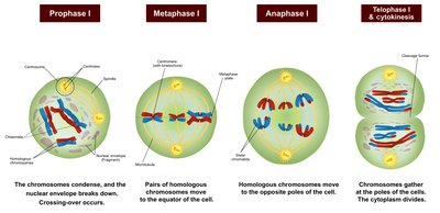
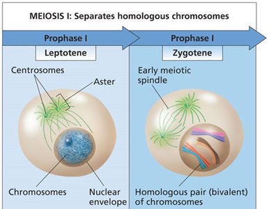
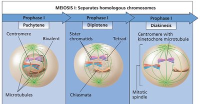
Prophase I Substages
Prophase I is subdivided into leptotene, zygotene, pachytene, diplotene, and diakinesis, each marked by specific chromosomal events.
Leptotene: Chromosomes condense.
Zygotene: Homologous chromosomes pair (synapsis).
Pachytene: Crossing over occurs.
Diplotene: Chiasmata visible, homologs begin to separate.
Diakinesis: Chromosomes fully condensed, spindle forms.
Genetic Variation in Meiosis
Meiosis introduces genetic variation through crossing over and independent assortment.
Crossing over: Exchange of genetic material between homologous chromosomes.
Independent assortment: Random distribution of homologs to gametes.
Comparison of Mitosis and Meiosis
Key Differences
Mitosis and meiosis differ in purpose, mechanics, and outcomes. Mitosis produces identical diploid cells, while meiosis generates genetically diverse haploid gametes.
Characteristic | Mitosis | Meiosis |
|---|---|---|
Purpose | Growth, maintenance | Sexual reproduction |
Location | Somatic cells | Germ-line cells |
Mechanics | One division | Two divisions |
Product | Two diploid cells | Four haploid cells |
Genetic variation | None | High (crossing over, assortment) |
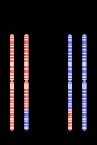
Chromosome Theory of Heredity
Genes Are Carried on Chromosomes
The chromosome theory of heredity, proposed by Sutton and Boveri, states that genes are located on chromosomes and their behavior during meiosis explains Mendel’s laws.
Mendel’s laws: Segregation and independent assortment are explained by chromosome movement.
Experimental evidence: Morgan’s studies in Drosophila melanogaster confirmed chromosomal inheritance.
Sex-Linked Inheritance
X-Linked and Y-Linked Traits
Sex-linked inheritance involves genes located on sex chromosomes. X-linked traits are inherited differently in males and females due to hemizygosity in males.
X-linked dominant: Expressed in heterozygous females and hemizygous males.
X-linked recessive: More frequently expressed in males.
Y-linked: Transmitted exclusively from father to son.
Human X-Linked Disorders
Several human diseases are caused by mutations in X-linked genes.
Disease | Symptoms |
|---|---|
Color blindness (red–green) | Color-perception deficiency |
Hemophilia A | Blood-clotting abnormality |
Fragile X syndrome | Mental retardation, neurodevelopmental defects |
Muscular dystrophy | Progressive muscle weakness |
Ornithine transcarbamylase deficiency | Mental deterioration due to ammonia accumulation |
Sex Determination
Chromosomal and Genetic Mechanisms
Sex determination is governed by the number and type of sex chromosomes. The X/A ratio (X chromosomes to sets of autosomes) determines gender in some species.
Females: Two X chromosomes (X/A ratio = 1.0)
Males: One X chromosome (X/A ratio = 0.5)
Dosage Compensation
Equalizing Expression of Sex-Linked Genes
Dosage compensation mechanisms balance gene expression between sexes. In placental mammals, random X-chromosome inactivation occurs in females, resulting in mosaicism.
Fruit fly: Male X-linked gene expression is doubled.
Roundworm: Hermaphrodite X expression is halved.
Marsupial mammals: Paternal X is inactivated in females.
Placental mammals: Random X inactivation in females.
Mechanism of X Inactivation
X inactivation is initiated by the XIST gene, which coats the X chromosome and silences its gene expression.
XIST gene: Produces RNA that inactivates one X chromosome.
Mosaicism: Results in visible phenotypic variation (e.g., calico cats).
Formulas and Equations
Chromosome Number in Mitosis and Meiosis
Mitosis:
Meiosis:
X/A Ratio
X/A Ratio:
