 Back
BackCell Division and Chromosome Heredity: Study Notes for Genetics Students
Study Guide - Smart Notes
 Tailored notes based on your materials, expanded with key definitions, examples, and context.
Cell Division and Chromosome Heredity
Introduction to Cell Division
Cell division is a fundamental process in genetics, enabling growth, maintenance, and reproduction in all living organisms. The cell cycle, which governs cell division, is highly conserved across eukaryotes and shares similarities with prokaryotic mechanisms, providing evidence for evolutionary relationships among all life forms.
Cell Cycle: The life cycle of a cell, including DNA replication and division.
Evolutionary Conservation: Many cell cycle genes are shared among plants, animals, and bacteria.
Importance: Essential for organismal growth, tissue repair, and genetic continuity.
Types of Cell Division
There are two main types of cell division: mitosis and meiosis. Each serves distinct biological purposes and results in different genetic outcomes.
Mitosis: Produces somatic cells for growth and repair. Results in two genetically identical daughter cells, maintaining the diploid chromosome number (2n).
Meiosis: Produces gametes (sperm and egg) from germ-line cells. Results in four genetically unique daughter cells, each with half the chromosome number (n).
The Cell Cycle
Phases of the Cell Cycle
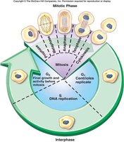
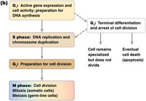
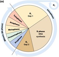
The cell cycle consists of two principal phases: Interphase and M phase. Interphase is subdivided into G1, S, and G2, while M phase includes mitosis or meiosis and cytokinesis.
G1 (Gap 1): Cell growth and gene expression.
S (Synthesis): DNA replication and chromosome duplication.
G2 (Gap 2): Preparation for cell division.
M phase: Cell division (mitosis or meiosis).
Interphase Details
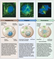
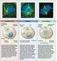
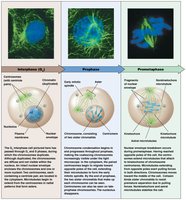
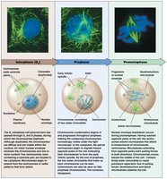
Interphase is the period of cell growth and DNA replication, preparing the cell for division. Chromosomes are duplicated but not yet visible, and centrosomes are replicated.
G1: Active gene expression and cell activity.
S: DNA replication.
G2: Preparation for mitosis or meiosis.
Chromosome Structure and Terminology
Chromosomes, Chromatids, and Sister Chromatids
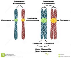
Understanding chromosome structure is essential for grasping cell division mechanics. Chromosomes are DNA-containing structures with a centromere. After DNA replication, each chromosome consists of two identical sister chromatids joined at the centromere.
Chromosome: DNA structure with a centromere.
Chromatid: Each DNA molecule in a replicated chromosome.
Sister Chromatids: Two identical chromatids joined by a centromere.
Mitosis: Phases and Mechanisms
Overview of Mitosis
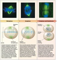
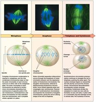
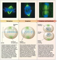
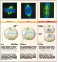
Mitosis is divided into five distinct phases: prophase, prometaphase, metaphase, anaphase, and telophase. Each phase is characterized by specific structural and molecular events.
Prophase: Chromosome condensation begins, centrosomes migrate, and spindle fibers start to form.
Prometaphase: Nuclear envelope breaks down, microtubules attach to kinetochores, chromosomes move toward the cell center.
Metaphase: Chromosomes align at the metaphase plate, spindle fibers fully formed.
Anaphase: Sister chromatids separate and move to opposite poles.
Telophase: Chromosomes decondense, nuclear envelope reforms, cytokinesis begins.
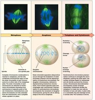
Chromosome Condensation and Alignment
Chromosomes condense during prophase and reach maximum condensation at metaphase. The centromere is the site of kinetochore formation, which attaches spindle fibers for chromosome movement.
Centromere: Specialized DNA sequence joining sister chromatids.
Kinetochore: Protein complex at the centromere for spindle fiber attachment.
Centrosomes and Microtubules
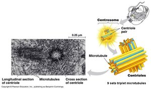
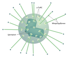
Centrosomes are the source of spindle fiber microtubules, which play critical roles in chromosome movement and cell stability during division. Microtubules have distinct plus and minus ends, and their organization forms the mitotic spindle.
Centrosome: Organelle containing two centrioles, source of microtubules.
Microtubules: Tubular structures with polarity, essential for spindle formation.
Types of Microtubules in Mitosis
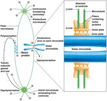
Three types of microtubules are involved in mitosis, each with specific functions:
Kinetochore microtubules: Attach to kinetochores and move chromosomes.
Polar microtubules: Extend toward opposite poles, elongate the cell.
Astral microtubules: Attach to the cell membrane, stabilize the cell.
Sister Chromatid Cohesion
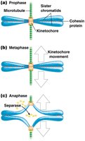
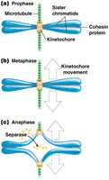
Cohesin proteins hold sister chromatids together, preventing premature separation. During anaphase, separase enzyme cleaves cohesin, allowing chromatids to separate.
Cohesin: Protein complex maintaining sister chromatid cohesion.
Separase: Enzyme that cleaves cohesin during anaphase.
Cytokinesis
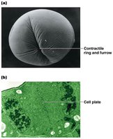
Cytokinesis is the final step of cell division, partitioning the cytoplasm and organelles between daughter cells. The mechanism differs between animal and plant cells.
Animal cells: Contractile ring of actin forms a cleavage furrow, pinching the cell.
Plant cells: Formation of a new cell wall along the midline.
Cell Cycle Regulation and Checkpoints
Cell Cycle Checkpoints
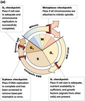
Cell cycle progression is tightly regulated by checkpoints, which ensure readiness for the next stage. These checkpoints are monitored by protein interactions and are crucial for maintaining genomic integrity.
G1 checkpoint: Checks cell size, nutrients, and growth factors.
S checkpoint: Monitors DNA replication and repair.
G2 checkpoint: Ensures DNA replication is complete and accurate.
Metaphase checkpoint: Verifies chromosome attachment to spindle fibers.
Cyclins and Cyclin-Dependent Kinases (Cdks)
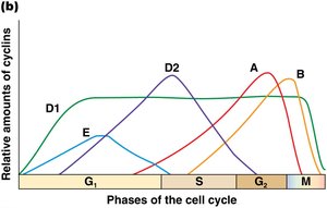
Cyclins and Cdks are protein complexes that drive cell cycle progression. Cyclins activate Cdks, which phosphorylate target proteins to regulate cell cycle transitions.
Cyclins: Regulatory proteins whose levels fluctuate during the cell cycle.
Cdk (Cyclin-dependent kinase): Protein kinase activated by cyclin binding.
Function: Control cell cycle transitions, activate/deactivate transcription factors, proteins, and signaling cascades.
Cell Cycle Mutations and Cancer
Mutations in cell cycle regulatory genes can lead to uncontrolled cell proliferation, a hallmark of cancer. Normal cells respond to growth factors and neighboring cells, while cancer cells ignore these signals, leading to tumor formation.
Normal cells: Proliferate only when needed, regulated by growth factors.
Cancer cells: Exhibit unregulated growth, invade and displace normal cells.
Summary Table: Cell Cycle Phases and Key Events
Phase | Main Event | Key Proteins/Structures |
|---|---|---|
G1 | Cell growth, gene expression | Cyclins, Cdks |
S | DNA replication | DNA polymerase, cohesin |
G2 | Preparation for division | Cyclins, Cdks |
Mitosis | Chromosome segregation | Spindle fibers, kinetochores, centrosomes |
Cytokinesis | Cytoplasm division | Actin (animals), cell wall (plants) |
Key Equations and Concepts
Diploid Number:
Haploid Number:
Chromatid Number After S Phase:
Conclusion
Understanding cell division and chromosome heredity is essential for genetics students, as these processes underpin genetic inheritance, organismal development, and the maintenance of genomic integrity. Mastery of the cell cycle, mitosis, and regulatory mechanisms provides a foundation for advanced study in genetics and molecular biology.
