 Back
BackCell Division: Mitosis and Meiosis – Genetics Study Notes
Study Guide - Smart Notes
 Tailored notes based on your materials, expanded with key definitions, examples, and context.
Cell Division in Genetics
Introduction to Cell Division
Cell division is a fundamental process in genetics, ensuring the transmission of genetic information from one generation to the next. It occurs through two main mechanisms: mitosis and meiosis. Mitosis produces genetically identical diploid cells, while meiosis generates haploid gametes, introducing genetic variation.
Chromosomes and Chromatin Structure
Chromatin Organization
In eukaryotic cells, DNA is packaged into chromatin, which condenses to form visible chromosomes during cell division. Chromatin fibers undergo multiple levels of folding and compaction, resulting in highly organized chromosomes.
Chromatin: Dispersed DNA-protein complex during interphase.
Chromosome: Condensed chromatin visible during mitosis/meiosis.
Centromere: Heterochromatic region determining chromosome shape and segregation.
Homologous Chromosomes
Diploid organisms possess pairs of homologous chromosomes, each carrying genes for the same traits but possibly different alleles. Homologs are similar in length and centromere position but are not identical.
Biparental inheritance: One chromosome of each pair from each parent.
Karyotype: The complete set of chromosomes in a cell, arranged by size and centromere position.
Sex chromosomes: X and Y in humans; not homologous in size or genetic content.
The Cell Cycle
Phases of the Cell Cycle
The cell cycle consists of a series of phases that prepare a cell for division. It includes interphase (G1, S, G2) and mitosis (prophase, prometaphase, metaphase, anaphase, telophase).
G1 phase: Cell growth and preparation for DNA synthesis.
S phase: DNA replication; chromosome number doubles.
G2 phase: Further growth and preparation for mitosis.
G0 phase: Quiescent state; cell is metabolically active but not dividing.
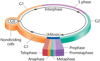
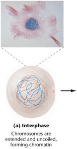
Interphase
During interphase, chromosomes are extended and uncoiled, forming chromatin. This phase is characterized by intense metabolic activity, cell growth, and differentiation.
Mitosis: Stages and Mechanisms
Overview of Mitosis
Mitosis is the process by which somatic cells divide, producing two genetically identical diploid daughter cells. It is essential for growth, development, tissue repair, and asexual reproduction.
Karyokinesis: Division of the nucleus.
Cytokinesis: Division of the cytoplasm and organelles.
Stages of Mitosis
Prophase: Chromatin condenses into chromosomes; nuclear envelope breaks down; spindle fibers form.
Prometaphase: Chromosomes attach to spindle fibers via kinetochores; centrioles reach poles.
Metaphase: Chromosomes align at the metaphase plate; spindle fibers fully attached.
Anaphase: Sister chromatids separate and migrate to opposite poles.
Telophase: Chromosomes decondense; nuclear envelope reforms; cytokinesis completes cell division.

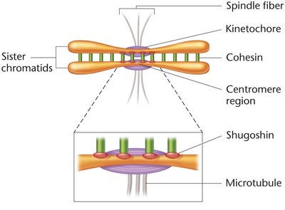
Key Molecular Components
Kinetochore: Protein structure on centromere for spindle attachment.
Cohesin: Protein complex holding sister chromatids together; degraded by separase during anaphase.
Shugoshin: Protects cohesin at centromeres until proper separation.
Meiosis: Generating Genetic Diversity
Overview of Meiosis
Meiosis is a specialized cell division that produces haploid gametes or spores, essential for sexual reproduction. It consists of two sequential divisions: meiosis I (reductional) and meiosis II (equational).
Meiosis I: Homologous chromosomes separate, reducing chromosome number by half.
Meiosis II: Sister chromatids separate, similar to mitosis.
Genetic variation: Achieved through independent assortment and crossing over.
Stages of Meiosis I
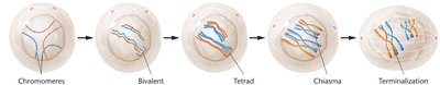
Prophase I: Homologous chromosomes pair (synapsis), forming bivalents and tetrads; crossing over occurs at chiasmata.
Metaphase I: Tetrads align randomly on the metaphase plate.
Anaphase I: Homologs separate; reductional division.
Telophase I: Two haploid cells form, each with dyads.
Stages of Meiosis II
Prophase II: Dyads (sister chromatids) prepare for division.
Metaphase II: Chromosomes align at the metaphase plate.
Anaphase II: Sister chromatids separate (equational division).
Telophase II: Four haploid gametes or spores are produced.
Genetic Variation in Meiosis
Independent assortment: Random alignment of homologs during metaphase I.
Crossing over: Exchange of genetic material between non-sister chromatids at chiasmata, resulting in recombinant chromosomes.

Gametogenesis: Spermatogenesis and Oogenesis
Spermatogenesis
Spermatogenesis is the process by which male gametes (sperm) are produced through meiosis. Each primary spermatocyte yields four haploid sperm cells, each with equal genetic content and cytoplasm.
Primary spermatocyte: Diploid cell entering meiosis.
Secondary spermatocyte: Haploid cell after meiosis I.
Spermatid: Haploid cell after meiosis II, matures into spermatozoa.
Oogenesis
Oogenesis is the process by which female gametes (eggs) are produced. It results in one large ovum and smaller polar bodies due to unequal cytoplasmic division.
Primary oocyte: Diploid cell entering meiosis.
Secondary oocyte: Haploid cell after meiosis I.
Ootid: Haploid cell after meiosis II, matures into ovum.
Polar bodies: Small cells with minimal cytoplasm, typically degenerate.
Major Events and Outcomes of Cell Division
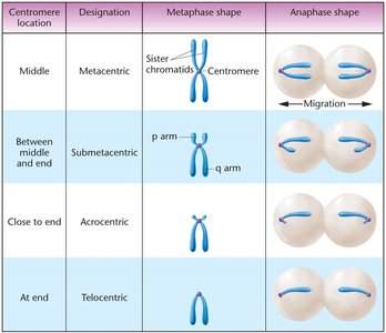
Summary Table: Chromosome Types by Centromere Location
The position of the centromere determines the classification and behavior of chromosomes during cell division.
Centromere location | Designation | Metaphase shape | Anaphase shape |
|---|---|---|---|
Middle | Metacentric | Sister chromatids, centromere in center | Migration to poles, V-shape |
Between middle and end | Submetacentric | p arm (short), q arm (long) | Migration to poles, L-shape |
Close to end | Acrocentric | Centromere near end | Migration to poles, J-shape |
At end | Telocentric | Centromere at tip | Migration to poles, I-shape |
Key Equations
Chromatid number after DNA replication:
Chromosome number after mitosis:
Chromosome number after meiosis:
Additional Info
Nondisjunction: Failure of homologous chromosomes or sister chromatids to separate properly, leading to aneuploidy.
Cell cycle checkpoints: Regulatory molecules ensure proper progression through cell cycle stages.
Genetic continuity: Achieved through accurate DNA replication and chromosome segregation.
